The exquisite detail provided by brain magnetic resonance imaging scans can make interpretation simultaneously straightforward and complicated, particularly to the novice. For this reason, it is essential to become familiar with normal structures before describing the pathologic condition. This article serves as a practical reference point to further enhance knowledge of the intracranial anatomy.
Brain magnetic resonance (MR) imaging studies provide multiple different imaging sequences in at least 2, and often 3, imaging planes. The different tissue signal characteristics and anatomic viewpoints are often complementary, and interpreting an MR imaging study of the brain can be a daunting task. The variety of pulse sequences and imaging planes makes understanding normal anatomy a necessity. Admittedly, many different approaches can be taken when interpreting images, but in this article just one approach to understanding normal anatomy is described. The normal anatomy of the brain has filled many textbooks, and a sincere effort has been made to provide a pertinent and concise reference suitable for review. Textbooks cited within the article serve as excellent references for those who wish to further their knowledge of brain anatomy.
Protocol
The overwhelming advantage of MR imaging is its ability to provide images with increased signal to noise ratios. Tissue characteristics with respect to different imaging sequences provide valuable clues when interpreting an MR image of the brain. Therefore, it is important to understand the accentuated tissue features on each scan. T1-weighted images provide good tissue discrimination and, in conjunction with the postcontrast scans, allow assessment for tissue enhancement. Precontrast and postcontrast scans must be obtained with identical imaging parameters to truly assess contrast enhancement ( Table 1 ). T1-weighted and T2-weighted images are complementary to each other because the T2-weighted images are sensitive to increased water content within tissues and to differences in susceptibility between tissues. Gradient echo images are susceptible to inhomogeneities in the magnetic field, accentuating blood products, iron, calcium, and manganese within tissues. This sequence is routinely performed during evaluation for stroke and trauma because hemorrhage is well seen. Fluid-attenuated inversion recovery (FLAIR) sequence can be used to obtain T2-weighted contrast while voiding the signal from cerebrospinal fluid, allowing a pathologic process to be identified with more confidence. Diffusion-weighted (DW) imaging assesses for the ability of water to diffuse in the local cell environment, which is particularly important in stroke and tumor imaging because areas of restricted diffusion demonstrate increased signal intensity. It is paramount to compare the DW images with the source apparent diffusion coefficient (ADC) images. Areas of restricted diffusion appear dark on the ADC maps, whereas areas of facilitated diffusion appear bright. Susceptibility-weighted images highlight differences in inherent tissue magnetic susceptibility and can be used for evaluation of deoxyhemoglobin in veins, hemorrhage, iron-containing tissues, and calcium deposition.
| Scan | Repetition Time (ms) | Echo Time (ms) | Inversion Time (ms) | Flip Angle (degrees) |
|---|---|---|---|---|
| 1.5-T Brain MR Imaging | ||||
| Sagittal T1 spin echo | 450 | 10 | NA | 90 |
| Axial T2 turbo spin echo | 5490 | 73 | NA | 150 |
| Axial gradient echo | 800 | 26 | NA | 20 |
| Axial FLAIR | 8900 | 141 | 2500 | 180 |
| Axial diffusion echo planar spin echo | 4000 | 83 | NA | NA |
| Axial T1 precontrast spin echo | 500 | 17 | NA | 90 |
| Axial T1 postcontrast spin echo | 500 | 17 | NA | 90 |
| Coronal T1 postcontrast spin echo | 500 | 17 | NA | 90 |
| 3.0-T Brain MR Imaging | ||||
| Sagittal T1 FLAIR | 2100 | 9 | 896.2 | 150 |
| Axial FLAIR | 9000 | 119 | 2500 | 150 |
| Axial T1 precontrast spin echo | 700 | 12 | NA | 90 |
| Axial T2 gradient echo | 700 | 19.9 | NA | 20 |
| Axial T2 susceptibility weighted | 27 | 20 | NA | 15 |
| Axial T2 turbo spin echo | 5440 | 96 | NA | 150 |
| Axial diffusion echo planar spin echo | 6400 | 109 | NA | NA |
| Axial T1 postcontrast spin echo | 700 | 12 | NA | 90 |
| Coronal T1 postcontrast spin echo | 700 | 8.9 | NA | 90 |
Identifying the lobes of the brain
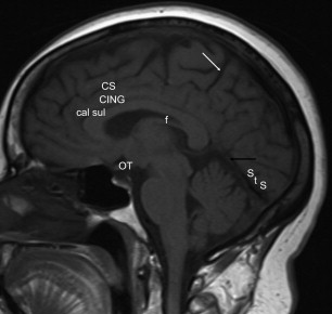
The frontal lobes are located anteriorly and extend posteriorly to the central (rolandic) sulcus, which partitions the frontal and parietal lobes. Several techniques can be used to identify the central sulcus, a universal point of reference. On the axial T2-weighted images near the vertex, the central sulci can be seen as a pair of mirror image transverse grooves ( Fig. 1 ), with the motor cortex always located anterior to this sulcus. The superior frontal sulcus is a horizontally oriented sulcus that terminates in the obliquely oriented precentral sulcus, and one can find the central sulcus as the sulcus posterior to the precentral sulcus. The precentral knob, the cortical location for hand function, is identified sitting just anterior to the central sulcus ( Fig. 2 ). The inferior central sulcus does not intersect the sylvian (lateral) fissure, rather it is contained by the junction of the precentral and postcentral gyri. On midline sagittal MR images, the central sulcus is somewhat more difficult to identify, but it is located anterior to the marginal ramus of the cingulate sulcus ( Fig. 3 ).


Inferiorly, the frontal lobe is separated from the temporal lobe by the sylvian fissure, which is easily seen on both axial and sagittal images ( Fig. 4 ). The middle cerebral arteries are located within the sylvian fissure and are seen as flow voids on T2-weighted images ( Fig. 5 ). The parietal lobes are bound anteriorly by the central sulcus. Superficially, there is no anatomic landmark separating the parietal and occipital lobes. However, toward the midline, the parietooccipital sulcus is well seen ( Fig. 6 ), demarcating their boundary.
Superficial Surface Anatomy
Although there are variations of normal anatomy, the superficial surface of the brain follows a general pattern, identified best on sagittal images. Naidaich and colleagues provide a thorough description of the superficial frontal and temporal lobes. The frontal lobe contains 3 horizontal gyri and the obliquely oriented precentral gyrus. The superior frontal gyrus runs horizontally, parallel to the falx and interhemispheric fissure. The middle frontal gyrus is the largest of the horizontal gyri, running parallel to the superior frontal gyrus and undulating posteriorly, where it fuses with the precentral gyrus. The superior frontal sulcus divides the superior frontal gyrus and middle frontal gyrus, and the inferior frontal sulcus divides the middle frontal gyrus and inferior frontal gyrus. The inferior frontal gyrus is triangular and is separated from the frontal pole by the frontomarginal sulcus. The superior border of the inferior frontal gyrus is horizontal, whereas the inferior surface is triangular and divided into 3 parts: the pars orbitalis, pars triangularis, and pars opercularis.
The superficial temporal lobe contains 3 superficial gyri: the superior temporal gyrus, middle temporal gyrus, and inferior temporal gyrus. The superior temporal sulcus separates the superior and middle temporal gyri and courses parallel with the superior fontal gyrus until posteriorly, where it angles superiorly and is called the angular sulcus. The inferior temporal sulcus separates the middle temporal and inferior temporal gyri.
The postcentral gyrus demarcates the anterior border of the parietal lobe and is parallel to the precentral sulcus. The intraparietal sulcus divides the parietal lobe into superior and inferior lobes. The inferior lobe is divided into the supramarginal gyrus and the appropriately named angular gyrus, which envelopes the angular sulcus and represents the posterior aspect of the inferior parietal lobe. The inferior parietal lobe can show marked left to right asymmetry.
The occipital pole can be seen along the posterior superficial surface, but most occipital lobe structures are better seen on the midline sagittal views, discussed next.
Midline Structures
The easiest structure to identify on the midline sagittal image is the corpus callosum, consisting of the rostrum, genu, body, and splenium (see Fig. 6 ). The cingulate gyrus parallels the corpus callosum anteriorly until its marginal branch courses to the superior brain surface. The sulcus anterior to the marginal sulcus is named the central sulcus. Posteriorly, the isthmus of the cingulate gyrus is located between the splenium of the corpus callosum and the anterior calcarine sulcus and continues laterally as the parahippocampal gyrus where its superior border is demarcated by the hippocampal fissure. The hippocampal fissure continues above the body of the corpus callosum as the callosal sulcus (see Fig. 3 ). Anteriorly, the cingulate gyrus dives under the rostrum of the corpus callosum and becomes the subcallosal area. The posterior medial parietal lobe, or precuneus, is seen anterior to the parietooccipital sulcus. The calcarine sulcus divides the medial occipital lobe into the cuneus and lingual gyrus (see Fig. 6 ).
The lamina terminalis demarcates the anterior wall of the third ventricle and plays a role in cardiovascular and body fluid homeostasis.
The hippocampal formation, found in the medial temporal lobe, is composed of the subiculum, the dentate gyrus, the hippocampus, and their continuations around the corpus callosum and is best seen on coronal images. The parahippocampal gyrus forms the inferomedial border of the temporal lobe ( Fig. 7 ). The subiculum forms the medial and superior curvature of the parahippocampal gyrus and arcs into the hippocampal fissure. The hippocampal fissure is bordered superiorly by the dentate gyrus and inferiorly by the subiculum. The hippocampus forms a cap on the hippocampal fissure and bulges into the medial wall and floor of the temporal horn.
The fornix, a white matter tract that connects the subiculum and the mammillary bodies, originates in the subiculum where its axons travel laterally to form a thin layer of white matter along the inferomedial temporal horn. The fornix continues posterior to the undersurface of the splenium, and most fibers arch upward, anterior, and inferior to the splenium to form the crura of the fornices, ending in the mammillary bodies (see Fig. 2 ).
The amygdala is a gray matter structure located just lateral to the uncus and anterior to the temporal horn and remains attached to the putamen superiorly. The tail of the caudate nucleus terminates in the amygdala. The anterior commissure is a white matter tract located in the anterior wall of the third ventricle at the junction of the lamina terminalis and the rostrum of the corpus callosum that connects the 2 temporal lobes and is easily seen on MR images.
Deep Structures
The deep central cerebral structures located between the insula and the sagittal midline are referred to as the central core ( Fig. 8 ). This core contains, among others structures, the extreme, external, and internal capsules, the claustrum, the putamen, the globus pallidus, the caudate nucleus, the amygdala, the diencephalon, and thalamic structures. It is related to motor and sensory functions, emotion, endocrine integration, and cognition. All the information passing between the brainstem and cortex passes through fibers in the central core. Anteriorly, the central core gray matter consists of the caudate nucleus and, to a lesser extent, the lentiform nucleus (putamen and globus pallidus), whereas the white matter consists primarily of the anterior limb of the internal capsule. More posteriorly, at the level of the foramen of Monro, the internal capsule contributes most of the white matter, whereas the lentiform nucleus contributes most of the gray matter, with a lesser contribution from the caudate nucleus. At the posterior insular level, most of the gray matter contribution arises from the thalamus and the white matter from the posterior limb of the internal capsule.