and Zdeněk Fryšák1
(1)
Department of Internal Medicine III – Nephrology, Rheumatology and Endocrinology, Faculty of Medicine and Dentistry, Palacky University Olomouc and University Hospital Olomouc, Olomouc, Czech Republic
Keywords
Thyroid volumeColor flow Doppler sonography patternLong-to-short axis ratioHilus sign1.1 Essential Facts
The thyroid gland is composed of two lobes connected by a median isthmus.
40% of all patients have an accessory lobe—the pyramidal lobe, which is more or less developed. It extends from the isthmus toward the hyoid bone, in front of the thyroid cartilage.
The thyroid lies in front of and on the sides of the trachea. It is bounded posterolateral by the carotid space, and its anterior and lateral aspects are covered by the strap muscles and the sternocleidomastoid muscles. The posterior surfaces of the lobes are adjacent to the perivertebral space and on the left to the esophagus [1].
A thyroid lobe has the shape of the rotation ellipsoid. Method of Brunn is widely used to calculate thyroid volume (Tvol). Tvol is a sum of the two lobes each calculated according to the following formula: V = width × depth × length × 0.479; or, an optimized correction factor 0.52 can be used [2, 3].
The contribution of the thyroid isthmus to total gland volume should be ignored [2]. But if the isthmus is >1 cm, its volume should be included [3].
Caution! Differences in technique (e.g., the pressure applied with the transducer) and in estimation of thyroid anatomy (e.g., inclusion of the thyroid isthmus and estimation of capsule thickness) can produce interobserver errors in Tvol as high as 26% [3].
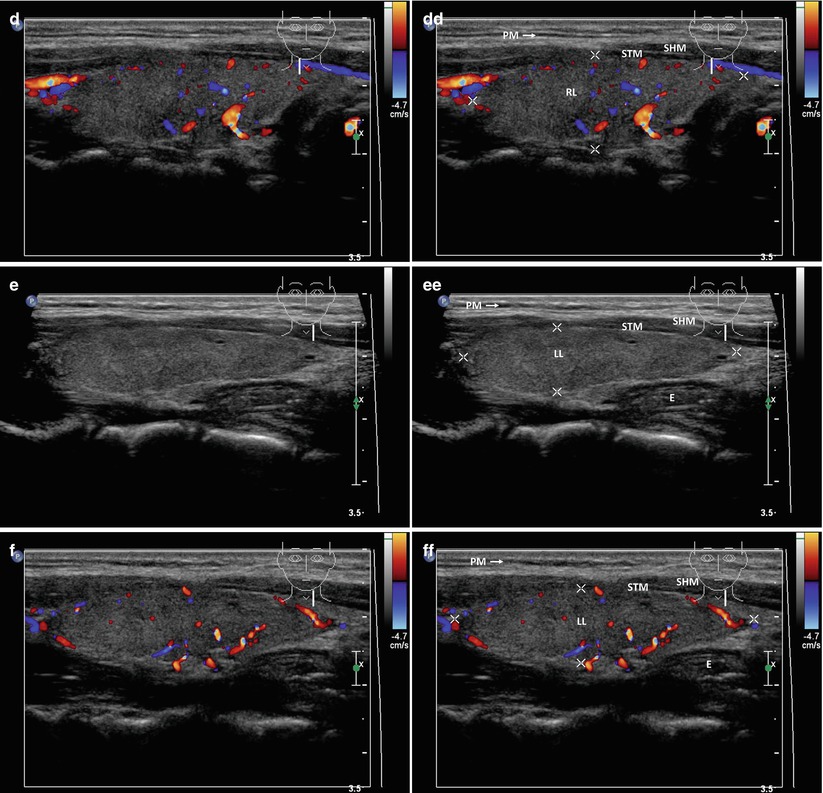
1.2 US Characteristics of the Thyroid Gland [4]
The echogenicity: isoechoic, hypoechoic, markedly hypoechoic, and hyperechoic patterns. The strap muscles and submandibular glands were used as a reference for the determination of echogenicity.
The echotexture: fine, coarse, and micronodulative patterns.
The glandular vascularity: normal, mildly increased, markedly increased, and decreased patterns.
The margin of the thyroid: smooth, microlobulated, and macrolobulated patterns.
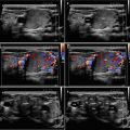
1.3 Color Flow Doppler Sonography (CFDS) Pattern [5]
Pattern 0 (Fig. 5.1dd): absent intraparenchymal (or nodular) vascularity or minimal spots.
Pattern I (Fig. 1.1bb): presence of parenchymal (or nodular) blood flow with patchy uneven distribution.
Pattern II (Fig. 3.15bb): mild increase of color flow Doppler signal with patchy distribution (for nodules: mainly peripheral).