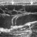
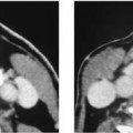
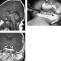
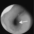
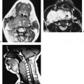
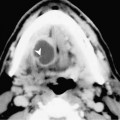
Chapter 156 Asymmetry of the internal jugular vein is not uncommon. One vein may be considerably larger than the other but this finding is of no clinical significance (Fig. 156–1). It is usually detected incidentally but patients may present clinically as having a vague neck mass. The clinician may also feel a soft mass in the neck. Occasionally, contrast may be seen layering in the dependent portion of the internal jugular vein (Fig. 156–1). This observation is of no clinical significance. It is not clear why layering should be present in an otherwise normal vein. Examination of the internal jugular vein fails to identify venous obstruction or possible causes of slow venous drainage. Rapid image acquisition using spiral CT often takes place before the various vascular structures show contrast opacification. The unopacified facial vein may mimic cervical nodes in the segment just before draining into the internal jugular vein (Fig. 156–2). Awareness of this potential pitfall should prevent a misdiagnosis. The unopacified facial vein can be traced to the internal jugular vein.
Normal Variants That May Mimic Disease
“Enlarged” Internal Jugular Veins
Contrast Layering in the Internal Jugular Vein
Unopacified Facial Vein Mimicking Lymphadenopathy
Stay updated, free articles. Join our Telegram channel

Full access? Get Clinical Tree


