The anatomic and histologic descriptions of the rotator cuff tendons and footprints are continuously evolving, and new discoveries have led to novel concepts in our understanding of rotator cuff tendon pathology. These concepts may be translated into the analysis of these footprints with imaging methods, particularly magnetic resonance imaging.
The anatomic and histologic descriptions of the rotator cuff tendons and footprints are continuously evolving, and new discoveries have led to novel concepts in our understanding of rotator cuff tendon pathology. These concepts may be translated into the analysis of these footprints with imaging methods, particularly magnetic resonance (MR) imaging.
Footprint of the supraspinatus, infraspinatus, and teres minor tendons
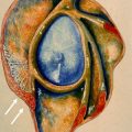
Understanding the osseous anatomy of the humeral head is an important requisite for describing the rotator cuff tendon insertions. The classic anatomy of the greater tuberosity of the proximal humerus describes 3 impressions or facets, which have often been designated as superior, middle, and inferior facets or, alternatively, as horizontal, oblique, and vertical facets ( Fig. 1 ). As in previous anatomic studies and traditional descriptions, these facets receive the insertional fibers of the supraspinatus, infraspinatus, and teres minor tendons, respectively. Some of the most extensive gross anatomic and histologic work was performed by Clark and Harryman. They found that the rotator cuff tendons fused as one inseparable structure at or near their insertions on the tuberosities of the humerus. The superficial fibers of the tendons generally maintained a parallel relationship with their respective muscle bellies. The supraspinatus and infraspinatus tendons were found to fuse into one inseparable tendon approximately 15 mm from their greater tuberosity insertions. These investigators depicted an interweaving of the supraspinatus and infraspinatus tendons as they coursed toward the greater tuberosity, with some of the anterior fibers of the infraspinatus tendon attaching far anteriorly ( Fig. 2 ). However, the actual anterior extent of these fibers was never elucidated.


In addition to their gross descriptions of the rotator cuff tendons, Clark and Harryman described 5 distinct histologic layers of the supraspinatus and infraspinatus tendons: (1) most superiorly, a thin (1 mm in thickness) superficial layer comprising fibers of the coracohumeral ligament; (2) a thicker layer (3–5 mm) of parallel tendon fibers; (3) a deeper layer (3 mm) comprising tendon fibers without uniform orientation, crossing over one another at 45° angles; (4) a layer of loose connective tissue with thick collagen bands, which merge with the coracohumeral ligament along the anterior edge of the supraspinatus tendon; and (5) a thin (1.5–2 mm) layer formed by the capsule of the joint, attaching to the greater tuberosity by the Sharpey fibers ( Fig. 3 ). In the third layer, the interdigitation of the supraspinatus and infraspinatus tendons resulted in their apparent fusion.

In a cadaveric study by Minagawa and colleagues, a similar pattern of overlap of the supraspinatus and infraspinatus tendons was described. However, these investigators described the supraspinatus tendon as the only tendon inserting on the superior facet of the greater tuberosity, with some fibers inserting on the superior half of the middle facet. The infraspinatus tendon covered the posterior half of the supraspinatus tendon and attached to the entire length of the middle facet. This type of interdigitation of the 2 tendons, which was described by Clark and Harryman and subsequently by Minagawa and colleagues, can be demonstrated on oblique sagittal images through the superior aspect of the cuff ( Fig. 4 ).

Clark and Harryman also described the existence of fibrous bands derived from the coracohumeral ligament that reinforced the superficial and deep portions of the supraspinatus and infraspinatus tendons near their greater tuberosity insertions. Regarding the relationship between the undersurface of the supraspinatus and infraspinatus tendons and the joint capsule, these structures adhered tightly near their humeral insertions. A 1-cm-wide thickening of the capsule derived from these fibrous bands of the coracohumeral ligament along the undersurface of the supraspinatus and infraspinatus tendons, located approximately 1.4 cm from the greater tuberosity footprint, was later termed the rotator cable , with the intervening portion of the cuff tendons designated as the crescent . The muscles, rather than the tendons, of the infraspinatus and teres minor were inseparable just proximal to their myotendinous junctions. The teres minor demonstrated a muscular insertion approximately 2 cm below its tendinous insertion on the greater tuberosity, a finding that can be demonstrated on MR images ( Fig. 5 ).

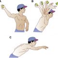
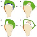
More recently, the concept of separate tuberosity attachments of the rotator cuff tendons has been reexamined, specifically regarding the shared footprint of the supraspinatus and infraspinatus tendons. In a cadaveric study, Mochizuki and colleagues revised the conventional understanding of this anatomy by demonstrating a relatively small, triangular, anteromedial footprint of the supraspinatus tendon at the superior facet and a comparatively large, trapezoidal footprint of the infraspinatus tendon occupying the anterolateral portion of the superior facet and the entire middle facet ( Fig. 6 ). Again, this configuration can be depicted on oblique sagittal MR images (see Fig. 4 ). This arrangement led the investigators to conclude that the development of infraspinatus muscle atrophy in the setting of apparently isolated supraspinatus tendon tear may be related to the far anterior insertion of the infraspinatus tendon and that the infraspinatus muscle may be more biomechanically important during shoulder abduction than previously thought ( Fig. 7 ).


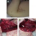
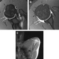
A possible related entity that has been recently described by Lunn and colleagues is that of a novel lesion of the infraspinatus characterized by musculotendinous disruption, edema, and late fatty infiltration ( Fig. 8 ). In their series of 19 patients, the investigators found full-thickness disruptions of the infraspinatus at the musculotendinous junction in 8 patients, within the tendon in 9 patients, and at an inconclusive site in 2 patients. These patients presented with acute pain, especially at night, with weakness of external rotation of the shoulder. This pathologic lesion was seen in middle-aged patients, with a mean age of 47.7 years, and more often in women than men. These investigators found a high incidence of accompanying rotator cuff pathology, including calcific tendinitis, tendinosis, and partial-thickness tears. Approximately 50% of their patients also reported a history of minor trauma, typically a fall on an outstretched arm. Only 1 of the 6 patients who underwent computed tomography (CT) arthrography demonstrated communication of contrast agent between the glenohumeral joint and the myotendinous junction of the infraspinatus.

Related posts:
 Internal Impingement Syndromes
 The Throwing Shoulder: the Orthopedist Perspective
 The Rotator Cable: Magnetic Resonance Evaluation and Clinical Correlation
 Magnetic Resonance Imaging of the Pediatric Shoulder
 Entrapment Neuropathies of the Shoulder
 Anatomic Variants and Pitfalls of the Labrum, Glenoid Cartilage, and Glenohumeral Ligaments
Stay updated, free articles. Join our Telegram channel

Full access? Get Clinical Tree


