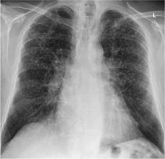
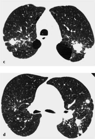
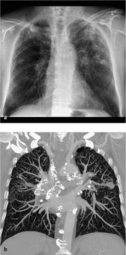
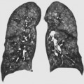
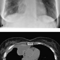
3 Occupational Diseases Mainly existing cases; occupational safety regulations have greatly reduced the incidence of new cases Alveolar phagocytosis and interstitial deposition of inhaled particles Radiographs, CT. Sharply demarcated, focal nodular lesions (1–10 mm in diameter) in the upper and middle lung fields, often with calcifications Micronodular lesions in the center and at the periphery of a lobule within the upper and middle lung fields Fig. 3.1 Anthracosilicosis in a 64-year-old man. Sharply demarcated, relatively dense, and partially confluent focal nodular lesions disseminated over both lungs, and increasing from base to apex. Bilateral apicolateral pleural calluses. Perihilar shadowing. Fig. 3.2 Anthracosilicosis in a 60-year-old man. The plain chest radiograph (a) and axial CT scans (c, d) show sharply demarcated nodules and large scarred focal lesions (conglomerates) in the upper lung fields, accompanied by shortening of the hila. CT also demonstrates bullous formations. The enlarged, severely calcified hilar and mediastinal lymph nodes, some exhibiting eggshell calcification, are best visualized on the coronal MIP (b). Symmetric micronodular changes in the upper and middle lung fields Dyspnea at rest and during exercise
Pneumoconiosis
Definition
 Epidemiology
 Chronic inhalation of inorganic dusts (especially silicate in crystalline form)
  Long-term exposure (sand blasters, quarry workers, miners).
 Etiology, pathophysiology, pathogenesis
 Development of interstitial reticulonodular granulomas of varying severity up to and including massive fibrosis.
  This leads to parenchymal shrinkage and scarring with emphysema.
Imaging Signs
 Modality of choice
 Radiographic findings
 These may become confluent, forming massive conglomerates
  Hilar and mediastinal lymph nodes show eggshell calcification.
 CT findings
 Signs of pulmonary fibrosis.
 Calluses.
Clinical Aspects
 Typical presentation
 Cough
Cough  Cyanosis
  Progressive fibrosis can lead to right heart failure.
 Therapeutic options
 Pathognomonic findings
Related posts:
Stay updated, free articles. Join our Telegram channel

Full access? Get Clinical Tree


