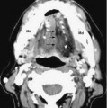
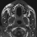
Functions
• Special afferent (SA) for sense of smell
Anatomy
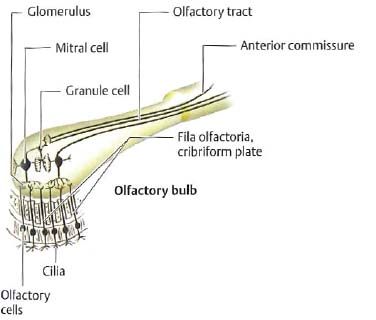
• The olfactory system (Fig. 1.1) consists of the olfactory epithelium, olfactory bulbs (Fig. 1.2), olfactory striae, and a variety of target brain areas partly though not exclusively devoted to processing olfactory information (Fig. 1.3).
• The olfactory nerves, like the optic nerves (II), differ from other cranial nerves in that they are really central nervous system (CNS) tracts. Both are composed of secondary sensory axons rather than primary sensory axons, and both have cellular constituents like the CNS and therefore demonstrate CNS rather than peripheral nervous system (PNS) pathologies (e.g., astrocytomas rather than schwannomas).
Olfactory Epithelium
• The olfactory epithelium has multiple cellular components, including the following:
 Olfactory cells. Bipolar neurons (~100 million) in the olfactory epithelium in the upper posterior nasal cavity. These cells have cilia projecting into mucus secreted by Bowman glands and express specific membrane receptor proteins to detect odorants (1 in Fig. 1.2).
 Sustentacular cells. Support the olfactory cells.
 Basal cells. The stem cells for new olfactory cells, which undergo constant replacement throughout life.
Olfactory Bulb and Tract
• Olfactory cells send bundles of unmyelinated axons (olfactory nerves proper) across the cribriform plate of the ethmoid bone to synapse on the second-order neurons in the olfactory bulb (Fig. 1.2).
• Mitral cells and tufted cells are the two types of secondary olfactory neurons in the olfactory bulb. At points of synapse with mitral and tufted cells, clusters of fibers called olfactory glomeruli are formed (Fig. 1.2). Granule cells (inhibitory interneurons) in the olfactory bulbs have no axons but form dendrodendritic synapses with mitral cells.
• The olfactory tract includes the axons of mitral and tufted cells and travels posteriorly in the olfactory sulcus between the gyrus rectus medially and the orbitofrontal gyrus laterally. It splits into lateral, medial, and intermediate olfactory striae at the anterior perforated substance.
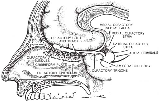
Fig. 1.1 Olfactory system (sagittal view). See text for details. (From Harnsberger HR. Handbook of Head and Neck Imaging (2nd ed.) St. Louis, MO: Mosby, 1995. Reprinted with permission.)
Olfactory Striae
• The olfactory trigone (Fig. 1.3) is the triangle formed by the lateral and medial striae.
• Lateral olfactory stria projects to the
 Anterior olfactory nucleus (Figs. 1.2, 1.3). Located between the olfactory bulb and tract, it receives fibers from the tufted cells and sends axons to either (1) cross in the anterior commissure to the contralateral anterior olfactory nucleus and olfactory bulb or (2) the ipsilateral olfactory cortical areas.
 Amygdala (Fig. 1.3).
 Primary olfactory cortex=the piriform (L., “pear-shaped”) cortex (Fig. 1.3) (lateral olfactory gyrus from lateral olfactory stria to amygdala) as well as the periamygdaloid cortex. This is the primary olfactory pathway.
• Fibers then travel from the piriform cortex (primary olfactory cortex) to the following:
 Entorhinal cortex (Fig. 1.3) (secondary olfactory cortex) to the hippocampus, insula, and frontal lobe by way of the uncinate fasciculus.
 Amygdala, lateral preoptic hypothalamus, and the nucleus of the diagonal band.
 Mediodorsal (MD) thalamic nucleus to the orbitofrontal cortex (for conscious analysis of odor, a phylogenetically newer pathway).
• Medial olfactory stria. Projects to the septal area (subcallosal area and the paraterminal gyrus) (also termed the medial olfactory area). This phylogenetically older pathway mediates the emotional/autonomic response to odors with its limbic connections.
• Intermediate olfactory stria. Projects to the anterior perforated substance (intermediate olfactory area) between the olfactory trigone (triangle formed by lateral and medial striae) and the optic tract.
• Rhinencephalon (“smell brain”). Olfactory bulbs, tracts, striae, and anterior olfactory nucleus and piriform cortex. Anterior perforated substance. Bounded by the medial and lateral olfactory striae anteriorly, optic tract medially, and posteriorly by the diagonal band of Broca. Transmits perforating vessels.
• Diagonal band of Broca. White matter tract connecting septal nuclei to amygdala, connects all three olfactory areas (medial, intermediate, and lateral).
• Efferent fibers from the olfactory areas travel in
• The medial forebrain bundle (MFB) from all three olfactory areas to the hypothalamus and brainstem reticular formation.
 The stria medullaris thalami from the olfactory areas to the habenular nucleus (epithalamus).
• The stria terminalis from the amygdala to the anterior hypothalamus and preoptic area. The hypothalamus sends the olfactory information to the reticular formation, superior and inferior salivatory nuclei (for salivation in response to odors), and dorsal motor nucleus of X (to cause acceleration of peristalsis and increased gastric secretion).
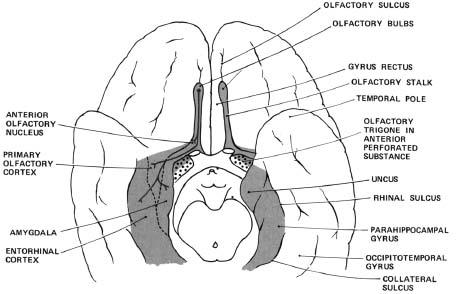
Fig. 1.3 Olfactory system (basal view). Together, the olfactory bulbs, tracts (“stalks”), striae, anterior olfactory nucleus, and piriform cortex make up the primary olfactory pathway. See text for details. (From Gilman S, Newman SW. Manter and Gatz’s Essentials of Clinical Neuroanatomy and Neurophysiology [10th ed.]. Philadelphia: F.A. Davis Publishers, 2003. Reprinted with permission.)
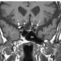
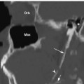
Olfactory Nerve: Normal Images (Figs. 1.4, 1.5, 1.6)
Olfactory System Lesions
• Loss of smell (anosmia) can be produced by a lesion anywhere along the olfactory pathway, from the olfactory epithelium, olfactory bulb, olfactory tracts, and/or central structures (posterior orbitofrontal, subcallosal, anterior temporal, or insular cortex).
•
Stay updated, free articles. Join our Telegram channel

Full access? Get Clinical Tree


