div style=”display:none;”>
Olfactory Nerve (CNI)
Main Text
TERMINOLOGY
Abbreviations
IMAGING ANATOMY
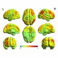
Overview
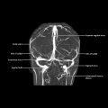
Nasal Epithelium
Intracranial Olfactory Bulb and Tract
 Histologically, bulb contains 6 concentric cell layers
 Axons within fila from receptor cells expressing same type of odorant receptor converge to spherical “glomerulus” in glomerular layer of bulb where they synapse with processes of secondary neurons (mitral and tufted cells) in deeper layers of bulb
 Axons of mitral and tufted cells coalesce to form lateral olfactory tract
 Recent studies have shown that main olfactory bulb is one of most prominent sites where intrinsic neurons are generated continuously after birth and in adulthood from cells located in subventricular zone of lateral ventricle
 This trifurcation creates olfactory trigone
 Anterior perforated substance is perforated by multiple small vascular structures
 Olfactory tract is made up of secondary sensory axons, not primary sensory axons
 Majority of fibers project through lateral olfactory stria and intermediate stria
 Anterior olfactory nucleus formed by some neurons along olfactory tract
 Olfactory tubercle is immediately behind division of olfactory stria, fused with anterior perforated substance