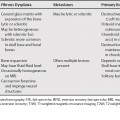
29 Vascular lesions represent a significant fraction of orbital masses and, as in the rest of the body, their characterization and classification are controversial, with frequent proposals for revision. Two of the more common lesions are varices and cavernous hemangiomas (Table 29.1). Varices are the most common venous malformation of the orbit, and represent local dilation of one or more abnormal venous channels. They have been subcategorized as distensible and nondistensible, based on the presence of expansion with increased venous pressure or lack thereof. They are also characterized as primary or secondary based on the presence of associated intraorbital or intracranial arteriovenous shunts. Some have classified these lesions, especially the primary and nondistensible lesions, along a spectrum of orbital venous abnormalities with lymphangiomas. They are unilateral, more often left-sided, and more commonly related to and parallel to the superior ophthalmic vein than the inferior ophthalmic vein. The radiographic triad of an enlarged orbit with venous lakes and phleboliths is rarely seen. They are intensely enhancing, well-defined round or elongated mass lesions within or outside the muscular cone that taper toward the apex and may only be evident with provocative maneuvers such as Valsalva. Their precontrast density and intensity depends on the presence of thrombus or hemorrhage.
Orbital Vascular Lesions
Orbital Varix
| Venous Varix | Cavernous Hemangioma | |
|---|---|---|
| Age | Before 4th decade | Middle age (mean = 43 years old) |
| Gender Predilection | None |


