Osteomyelitis and Discitis (Spondylodiscitis)


Clinical Presentation


The patient is a 39-year-old otherwise healthy woman who developed a papulopustular skin eruption/infection attributed to MRSA (methicillin-resistant Staphylococcus aureus ). One month later, she developed pleuritic chest pain and a cough and was diagnosed with pneumonia. The symptoms resolved, but pleuritic chest pain recurred along with back pain. The patient was afebrile. Magnetic resonance (MR) imaging of the thoracic spine was ordered.


Imaging Presentation


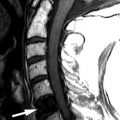
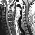
MR imaging of the thoracic spine revealed signal abnormalities consistent with T5-6 discitis and vertebral osteomyelitis ( Figs. 56-1 to 56-4 ) . A paraspinal phlegmon was also demonstrated ( Fig. 56-5 ) . A percutaneous paraspinal computed tomography (CT)–guided biopsy was performed at the T5-6 level, which yielded no viable organisms ( Fig. 56-6 ) . This was followed with a percutaneous transpedicular biopsy ( Fig. 56-7 ) . A diagnosis of MRSA osteomyelitis and discitis was made based on the culture of biopsy tissue.


Figure 56-1


Spondylodiscitis at T5-6 Level.

Sagittal T2-weighted MR image reveals partial collapse of the T5 and T6 vertebral bodies. Abnormal T2 hyperintensity visible throughout the T5 and T6 vertebral body marrow. The T5-6 vertebral endplates are indistinct and ill-defined tissue is located within the T5-6 intervertebral disc ( arrow ). T7 = T7 vertebral body.


Figure 56-2


Spondylodiscitis at T5-6 Level.

A prevertebral soft tissue mass ( short arrows ) is evident on this sagittal T1-weighted MR image in same slice location as in Figure 56-1 . The T5 and T6 vertebral marrow is relatively T1 hypointense compared to the marrow signal intensity in normal adjacent vertebrae. The T5-6 vertebral endplates are poorly defined and the intervertebral disc ( long arrow ) contains tissue that is slightly T1 hyperintense relative to adjacent vertebral bodies. T7 = T7 vertebral body.


Figure 56-3


Spondylodiscitis at T5-6 Level.

Same patient as in Figures 56-1 and 56-2 . Sagittal contrast-enhanced fat-saturated T1-weighted MR image. The prevertebral phlegmonous soft tissue mass ( short arrows ) enhances following IV contrast. Intense enhancement is seen throughout the T5 and T6 vertebral bodies. There is diffuse enhancement of inflammatory tissue in the T5-6 intervertebral disc and minimal enhancement of inflammatory tissue in the ventral epidural space ( long arrow ). T7 = T7 vertebral body.


Figure 56-4


Spondylodiscitis at T5-6 Level.

Axial contrast-enhanced fat-saturated T1-weighted MR image at the T6 level in same patient as in Figure 52-1 , Figure 52-2 , Figure 52-3 , shows contrast enhancement of the T6 vertebral body and paraspinal phlegm ( arrows ). A = aorta.


Figure 56-5


Spondylodiscitis at T5-6 Level.

Corona CT image obtained through thoracic vertebral bodies in same patient as in Figures 56-1 to 56-4 . Paraspinal soft tissue mass ( arrows ) represents paravertebral phlegmon, which extends from C5 to C7 level. Note partial collapse of vertebral body superiorly and increased bone density in C5 and C6 vertebral bodies. T5-6 vertebral endplates are slightly indistinct, irregular and sclerotic.


Figure 56-6


Spondylodiscitis at T5-6 Level, CT-Guided Biopsy.

Axial CT image obtained through C6 level with patient prone during CT guided percutaneous biopsy in same patient as in Figures 56-1 to 56-5 . The biopsy needle ( arrow ) is positioned in the right paraspinal soft tissue mass. A = aorta.


Figure 56-7


Spondylodiscitis at T5-6 Level, X-Ray Fluoroscopic-Guided Biopsy.

PA radiograph of the mid-thoracic spine with patient prone during percutaneous fluoroscopic-guided needle biopsy performed using right T6 transpedicular approach. The needle tip projects over the right T6 pedicle ( P ). T5 and T6 lettering overly the right T5 and T6 transverse processes, respectively.


Discussion


Spondylodiscitis is an infectious process involving the vertebra ( osteomyelitis ) and adjacent intervertebral disc ( discitis ). Rarely, tuberculosis and other granulomatous infections can cause vertebral infection without invading the intervertebral disc, but the vast majority of cases of osteomyelitis penetrate the vertebral endplate to involve the intervertebral disc. The vertebral-disc infection frequently extends into the paraspinal and epidural space forming a phlegmon, which may cavitate to form an abscess. Rarely, the infection infiltrates the thecal sac where it can cause arachnoiditis, meningitis, or an intramedullary spinal cord abscess.


Pyogenic and granulomatous spondylodiscitis occurs more frequently in patients with a chronic illness, including patients with diabetes mellitus, chronic renal failure, or chronic liver disease; immunocompromised patients; and IV drug abusers. Spondylodiscitis also occurs in patients with chronic nutritional deficiencies such as those in chronic alcoholics.


Most vertebral infections occur from blood-borne pathogens that preferentially settle in the hypervascular marrow adjacent to the vertebral endplates. In adults, the disc is an avascular structure and becomes secondarily involved after the infectious process destroys the vertebral endplates. The vertebral cortical and cartilaginous endplates represent the nutritional conduit for the intervertebral disc. When the cortical and cartilaginous endplate becomes disrupted, the infection can enter directly into the disc. In children, the intervertebral disc may still be vascularized, so the infectious process can begin primarily within the disc. Infections can also invade the vertebra by direct extension from infections in adjacent organs such as infections originating in the genitourinary or gastrointestinal tract. Infections may also occur in the vertebra after trauma, surgery, or percutaneous spine interventional procedures.


Vertebral infections are most commonly caused by pyogenic bacteria. The most common pathogen is Staphylococcus aureus (see Figs. 56-1 to 56-5 ). However, other organisms may cause pyogenic spine infections, including coagulase negative Staphylococcus ( Figs. 56-8 and 56-9 ) , Streptococcus, Pneumococcus, Hemophilus, Enterococcus, Escherchia coli, Salmonella, and rarely anaerobic organisms. Granulomatous spinal infections are seen with increasing frequency, especially in immunocompromised patients. Granulomatous spondylodiscitis is most commonly produced by Mycobacterium tuberculosis ( Fig. 56-10 ) . However, atypical mycobacteria ( Figs. 56-11 to 56-13 ) , Brucella, Streptomyces , parasites, and fungal organisms can also cause granulomatous spine infections. Fungal spondylodiscitis is a rare spinal infection, usually occurring in immunocompromised patients. Infections produced by both common and uncommon pathogens more frequently occur in immunocompromised patients.


Figure 56-8


L3-4 Discitis and Osteomyelitis Secondary to Coagulase Negative Staphylococcus .

Lateral radiograph of lumbar spine reveals obscure intervertebral disc space ( arrow ) and indistinct vertebral endplates, secondary to vertebral bone destruction on either side of L3-4 intervertebral disc. Reduction in vertebral body height of L3 and L4 is also demonstrated. Note narrowed L4-5 and L5-S1 intervertebral disc spaces associated with well-defined, sclerotic endplates, secondary to intervertebral disc degeneration.


Figure 56-9


L3-4 Discitis and Osteomyelitis.

Same patient as in Figure 56-8 . Sagittal fat-saturated T2-weighted MR image A . T1-weighted image B , and contrast-enhanced fat-saturated T1-weighted MR image C . The L3-4 intervertebral disc ( arrow in images A , B , and C ) is narrowed and adjacent vertebral endplates are irregular and enhance intensely. The L3-4 intervertebral disc contains T1 hypointense and heterogeneous T2 hyperintense tissue. On image C , intradiscal inflammatory tissue enhances but intradiscal fluid does not enhance. The L3 and L4 vertebra are T1 hypointense and T2 hyperintense relative to other vertebral bodies, due to presence of marrow edema. The L3 and L4 vertebral bodies enhance diffusely indicating presence of vertebral inflammation secondary to osteomyelitis.


Figure 56-10


C6-7 Spondylodiscitis Secondary to Mycobacterium Tuberculosis Infection.

On sagittal T1-weighted MR image A , the C6-7 intervertebral disc space has been replaced by heterogeneous tissue and adjacent endplates are indistinct. On sagittal gradient echo images, the C6-7 intervertebral disc space contains hyperintense tissue and adjacent vertebral endplates and vertebral bodies are destroyed. The MR appearance in this case of tuberculous spondylodiscitis is nonspecific and could be produced by almost any organism capable of producing discitis.


Figure 56-11


L2-3 Spondylodiscitis, Epidural and Paraspinal Abscesses Caused by Atypical Mycobacterium ( M. Abscessus ).

On sagittal T2-weighted MR image A , the L2-3 intervertebral disc is narrowed, undulating and contains ill-defined T2 hyperintense tissue ( arrows ). The posterior disc margin ( long arrow ) protrudes into the spinal canal. The anterior disc margin ( short arrow ) protrudes into the prevertebral tissues. Heterogeneous T2 hyperintense tissue is seen in the posterior paraspinal region from L1-3. On sagittal T1-weighted image B , the disc space appears widened, but in reality is not widened, due to presence of T1 hypointense tissue between T1 hypointense curvilinear areas of bony reactive response ( arrows ) in the L2 and L3 vertebral bodies. On sagittal contrast-enhanced fat-saturated T1 weighted MR image C , the upper and lower margins of the tissue within the L2-3 intervertebral disc display intense, uniform, curvilinear enhancement ( short arrows ). Enhancing epidural and posterior paraspinal tissues enhance intensely ( long arrows ), representing epidural and paraspinal inflammatory tissue (phlegmon).


Figure 56-12


L2-3 Spondylodiscitis, Epidural and Paraspinal Abscesses Caused by Atypical Mycobacterium.

Same patient as in Figure 56-11 . On this axial contrast-enhanced fat-saturated T1-weighted MRI, a large paravertebral phlegmon ( large arrows ) contains multiple small fluid collections representing multiple abscesses. The thecal sac is displaced to the right by epidural phlegmon ( small arrows ) which contains several small abscesses.


Figure 56-13


L2-3 Spondylodiscitis, Epidural and Paraspinal Abscesses Caused by Atypical Mycobacterium.

Same patient as in Figures 56-11 and 56-12 . Coronal contrast-enhanced fat-saturated T1-weighted MRI. Lumbar dextroscoliosis. The tissue at the superior and inferior margin of the L2-3 intervertebral disc ( small arrows ) enhances intensely due to discitis. Both psoas muscles enhance diffusely and contain non-enhancing fluid filled cavities ( long arrows ), which represent abscesses in the psoas muscles.


The diagnosis of spondylodiscitis should be made based on clinical information and the imaging appearance. Patients with spine infections typically present with acute or progressively worsening back pain. The pain tends to have an acute onset with pyogenic infections and a more insidious onset in granulomatous infections, but in both cases, the pain becomes progressively worse. Constitutional symptoms may be present including fever, chills, malaise, and night sweats. These patients may present with myelopathic symptoms if the cord is compressed by epidural phlegmon or abscess. If the phlegmon extends into the neural foramen or paraspinal soft tissues, they may have radicular symptoms.


Leukocytosis with a high neutrophil count, markedly elevated ESR (erythrocyte sedimentation rate), and C-reactive protein levels are strongly suggestive of a pyogenic infection. In granulomatous infections (e.g., tuberculosis, Brucella ), the ESR is usually elevated out of proportion to the degree of leukocytosis.


If a disc infection is suspected, it is important to perform percutaneous needle aspiration biopsy of the disc, adjacent vertebra, and/or phlegmonous tissue to culture the offending organism and determine the most effective antibiotic for treatment (see Figs. 56-6 and 56-7 ). However, one should not rely on a positive culture after aspiration biopsy to initiate therapy because the results are commonly negative for a variety of reasons. The disc aspiration may yield negative results if the patient has already received antibiotic therapy or if samples are taken only from the disc center, which may contain only noninfectious liquid and necrotic debris. It is important that aspiration samples be obtained from inflamed, contrast-enhancing tissue in the involved vertebral body, adjacent to a bony sequestrum or in a paravertebral phlegmon. Disc aspiration biopsy often yields negative results in patients with mycobacterial infections, so it is important to always consider tuberculosis as a possible cause of the infection.


It requires about 4 weeks to culture M. tuberculosis, so it is not practical to withhold therapy while waiting for culture results. However, the diagnosis of tuberculosis can also be made if acid-fast bacilli are detected in the tissue samples (using an enzyme-linked immunosorbent assay for tuberculous bacilli) or if characteristic Langhans giant cells are seen in the tissue. If tuberculosis is suspected, a positive Mantoux (purified protein derivative [PPD]) skin test supports the diagnosis of tuberculosis. A PCR (polymerase chain reaction) test should be performed on the biopsy tissue samples. PCR is a quick, reliable DNA test that is used to confirm the diagnosis of tuberculosis. However, the PCR may be, in a minority of cases, falsely negative, so a negative PCR does not exclude the diagnosis of tuberculous spondylitis. If caseating granulomas are observed histologically in the tissues, this indicates either tuberculosis or brucellosis.

Only gold members can continue reading. Log In or Register to continue

Stay updated, free articles. Join our Telegram channel

Aug 25, 2019 | Posted by in NEUROLOGICAL IMAGING | Comments Off on Osteomyelitis and Discitis (Spondylodiscitis)

Full access? Get Clinical Tree

Get Clinical Tree app for offline access