Osteosarcoma of H&N
H. Ric Harnsberger, MD
Key Facts
Terminology
Definition: Malignant tumor arising from bone with ability of neoplastic cells to produce osteoid
Imaging
Bone CT: Shows bone tumor with both osteolytic & osteoblastic components
MR imaging: Best evaluates extent of osteosarcoma
Intramedullary and extraosseous soft tissues
PET/CT: Used for local recurrence and distant metastasis identification
Top Differential Diagnoses
Mandible-maxilla osteomyelitis
Mandible-maxilla metastasis
Ewing sarcoma
Langerhans cell histiocytosis
Mandible-maxilla osteoradionecrosis
Pathology
Heterogeneous mass with ossified and nonossified components
Chondroblastic > osteoblastic > fibroblastic
Clinical Issues
Mean age: 35 years
Prognosis depends on pathologic type, size, location, and presence of metastases
5-year survival = 60%
Complete resection affords best chance of survival
Diagnostic Checklist
Osteoid matrix in tumor of mandible or maxilla suggests osteosarcoma
If not present, consider metastasis or osteomyelitis
Consider radiation-induced osteosarcoma if patient had radiation years prior
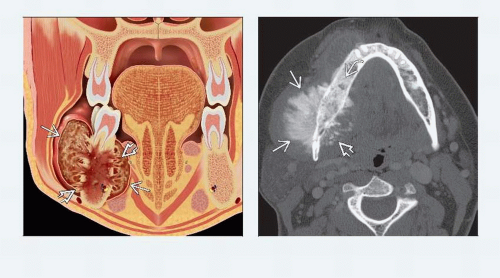 (Left) Coronal graphic shows right mandible osteosarcoma. Note associated soft tissue mass  with aggressive periosteal reaction with aggressive periosteal reaction  . (Right) Axial bone CT demonstrates large, dense mass arising from the right mandibular body. The mass has both osteoid matrix . (Right) Axial bone CT demonstrates large, dense mass arising from the right mandibular body. The mass has both osteoid matrix  and periosteal reaction and periosteal reaction  associated with it. This is a classic aggressive periosteal reaction associated with osteosarcoma where periosteum is lifted off perpendicular to bone. Marrow within the involved portion of the mandible is sclerotic associated with it. This is a classic aggressive periosteal reaction associated with osteosarcoma where periosteum is lifted off perpendicular to bone. Marrow within the involved portion of the mandible is sclerotic  . . |
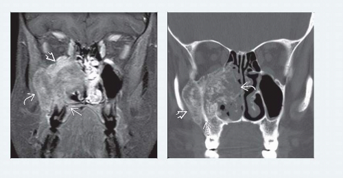 (Left) Coronal T1 C+ fat-saturated MR reveals a large, heterogeneously enhancing maxillary mass involving the alveolar ridge  , orbital floor , orbital floor  , and deep soft tissues of the cheek , and deep soft tissues of the cheek  . (Right) Coronal bone CT in the same patient shows extensive new bone formation in the matrix, which creates central low signal. The tumor is centered on the lateral wall of the right maxillary sinus . (Right) Coronal bone CT in the same patient shows extensive new bone formation in the matrix, which creates central low signal. The tumor is centered on the lateral wall of the right maxillary sinus  , extending into the fat pad , extending into the fat pad  and nasal cavity and nasal cavity  . Most maxillary OSa are found in the alveolar ridge, not the lateral wall as in this case. . Most maxillary OSa are found in the alveolar ridge, not the lateral wall as in this case. |
TERMINOLOGY
Abbreviations
Osteosarcoma of head & neck (OSa H&N)
Synonyms
Osteogenic sarcoma
Definitions
Malignant tumor arising from bone with ability of neoplastic cells to produce osteoid
IMAGING
General Features
Best diagnostic clue
H&N bone tumor demonstrating tumor matrix mineralization with aggressive bone destruction and soft tissue extension leads directly to radiologic diagnosis of osteosarcoma
Location
Mandible ≈ maxilla > > calvarium/skull base
All other sites are extremely rare
Hard palate, mastoid, zygoma, paranasal sinuses
Mandible OSa in mandibular body
Maxillary bone OSa in alveolar ridge
Postradiation OSa: Typically at border of radiation field
Most commonly involves multiple bones at this site
Size
Ranges in size from 1-15 cm
Majority present in 3-6 cm size range
Median size is 5.5 cmRelated posts:

Stay updated, free articles. Join our Telegram channel

Full access? Get Clinical Tree


