Fig. 1.1
The piezoelectric effect is a bidirectional phenomenon whereby an alternating current applied to a crystal creates vibration (panel a); additionally, oscillation of the crystal by sound waves generates a voltage across the crystal (panel b)
Detection of an Ultrasound Echo
The physical property that describes the ability of sound to move through an object is called acoustic impedance, which is a function of both density and speed of sound through a substance. When a sound wave reaches a boundary between two entities with different acoustic impedances, a portion of the wave is transmitted and the rest is reflected back to the source as an echo (Fig. 1.2). The degree to which a structure reflects sound pulses during an ultrasound examination is denoted as “echogenicity.” Highly echogenic, or hyperechoic, structures typically have relatively low water content, reflect a large proportion of incoming ultrasound, and are displayed as white on a B-mode image. Commonly seen hyperechoic tissues include cortical bone, tendon, organ and muscle sheaths, nerves, and gallstones. Extremely hyperechoic structures result in an artifact on the image known as acoustic shadowing; since most of the sound has been reflected to the probe at the structure’s surface, the area behind the echogenic material is displayed as a black streak (Fig. 1.3).
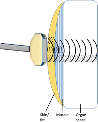
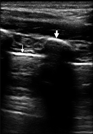
Fig. 1.2
A small portion of the beam created by an ultrasound probe is reflected at each boundary between substances with varying acoustic impedances. The echoed wave is detected by the same transducers within the probe and used to form an image for display
Fig. 1.3
Hyperechoic structures are seen on B-mode ultrasound as bright white. If the object is sufficiently opaque to sound, an acoustic shadow artifact is formed. In this image taken of the chest wall of a 6-year-old boy with a linear probe, both the cortical bone of the rib (thick arrow) and the lung pleura (thin arrow) appear hyperechoic. The rib does not transmit the ultrasound beam further, thus causing an acoustic shadow
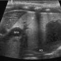
Features with low echogenicity are known as hypoechoic or anechoic and are seen on B-mode as dark grey or black, respectively. These include solid organs , muscle bellies, lymph nodes, vasculature, and other structures that are fluid filled or high in water content.
Formation of an Image
The simplest mode of ultrasound is known as A-mode (amplitude), in which a single piezoelectric transducer detects the distance to an echogenic structure by precisely measuring the time between emitting a pulse and receiving an echo, utilizing the equation distance=velocity * time. Thus, using the known speed of sound and the detected time to echo, the distance to an object can be calculated. A-mode itself is generally only of historic interest, but can be helpful to understand B-mode (brightness), which is the characteristic mode utilized in modern diagnostic ultrasound. In B-mode imaging, a row of transducers simultaneously act as just described, and the resultant echoes are compiled to produce a two-dimensional image. This is repeated at least 20 times per second to create a real-time effect. An important assumption in creating a B-mode image using the equation above is that the speed of sound through soft tissue is uniform, most frequently assumed to be a constant 1540 m/s, slightly higher than the speed of sound through pure water. However, in reality, the soft tissues encountered in medical ultrasound have a range of acoustic impedances and resultant velocities of sound, which can lead to imprecise localization of an object’s depth and is a limiting factor in the axial resolution of the modality in general.
Other commonly employed ultrasound modes are M-mode (movement) and Doppler imaging. M-mode is used to track the movement of an object over time. Initially, a two-dimensional B-mode image is acquired and a scan line is placed along the area of interest. The movement over time of each echogenic interface intersected by that line will then be displayed. M-mode is ideal for characterizing precise temporal events such as the motion of cardiac valves.
Related posts:


Stay updated, free articles. Join our Telegram channel

Full access? Get Clinical Tree


