• This encompasses a spectrum of cystic posterior fossa malformations from the complete Dandy–Walker malformation to a mega cisterna magna • It is associated with hydrocephalus and other midline abnormalities (e.g. agenesis or a lipoma of the corpus callosum) • Total aplasia of the cerebellar vermis reflecting the failure of formation of the decussation of the superior cerebellar peduncles, a lack of the pyramidal decussations, and other anomalies of the midbrain crossing tracts and their nuclei • Occasionally a genetic locus has been identified • Many syndromes with additional features (e.g. renal cysts, ocular abnormalities, liver fibrosis, hypothalamic hamartomas and polymicrogyria) have been classified with this anomaly • A developmental mass lesion with enlargement of the cerebellar cortex A non-enhancing mass with diffusely enlarged cerebellar folia (± pial enhancement) • A form of hindbrain deformation rather than a true malformation characterized by tonsillar descent • It is often an isolated hindbrain abnormality of little consequence • There are usually no symptoms during childhood unless there is an associated syringomyelia or hydrocephalus • Clinical symptoms are more likely when there is > 5mm of descent below the foramen magnum (children between 5 and 15 years can have normal tonsillar descent of up to 6mm) • Symptoms may include a cough-induced headache, cranial nerve palsies and a disassociated peripheral anaesthesia • A congenital malformation of the hindbrain (with a dysplastic cerebellum) that is almost always associated with a myelomeningocele • The inferior vermis is everted (rather than inverted) so that the nodulus becomes its most inferior aspect and the 4th ventricle is reduced to a coronal cleft (the cerebellar herniation consists mainly of the cerebellar vermis) • ‘Tectal beaking’: this follows fusion of the midbrain colliculi into a single beak pointing posteriorly • ‘Towering cerebellum’: the tentorial incisura is enlarged and the cerebellum herniates superiorly into the supratentorial space • ‘Batwing’ configuration of the frontal horns (coronal view): this is due to impressions from prominent caudate nuclei • ‘Hourglass ventricle’: a small biconcave 3rd ventricle due to a large massa intermedia • ‘Cervicomedullary kink’: herniation of the medulla posterior to the spinal cord • ‘Banana’ sign: the cerebellum is wrapped around the posterior brainstem (seen during obstetric US) • These are malformations related to the formation of the neural tube (also including Chiari II malformations) • An extracranial protrusion of intracranial structures through a congenital defect of the skull and dura mater • Meningocele: containing leptomeninges and CSF only • Encephalocele: containing leptomeninges, CSF and neural tissue • Encephalocystocele: containing leptomeninges, CSF, neural tissue and part of the ventricle • A midline malformation of ventral induction of the anterior brain, skull and face (resulting from the failure of the embryonic prosencephalon to undergo segmentation and cleavage into two separate cerebral hemispheres) • The anterior part (the posterior genu and anterior body) of the corpus callosum is formed before the posterior part (the posterior body and splenium) • This is located parallel to the ventricular wall and is seen as a homogeneous band of grey matter between the lateral ventricle and the cerebral cortex (separated from both by a layer of white matter) • It is commonly seen in girls with variable developmental delay or seizures
Paediatric neuroradiology
CEREBELLAR MALFORMATIONS
CEREBELLAR HYPOPLASIA
DEFINITION
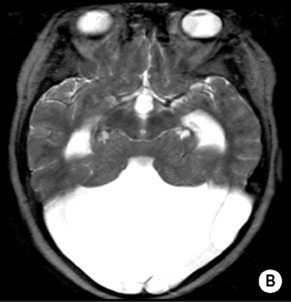
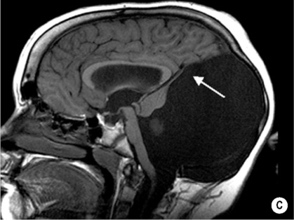
DANDY–WALKER COMPLEX
DEFINITION
 Membranous obstruction to the foramina of Magendie and Luschka causes cystic dilatation of the 4th ventricle
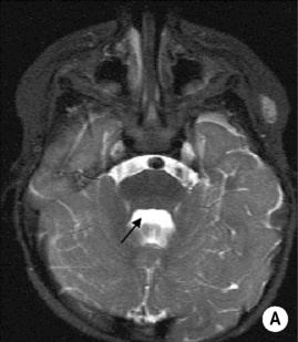
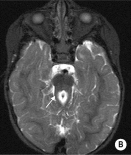
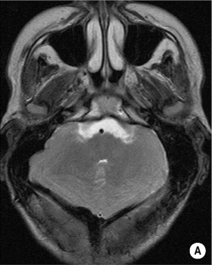
JOUBERT’S SYNDROME
DEFINITION
OTHER CEREBELLAR MALFORMATIONS
Lhermitte–Duclos or dysplastic cerebellar gangliocytoma
 this usually affects one hemisphere
MRI
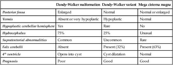
Dandy–Walker malformation
Dandy–Walker variant
Mega cisterna magna
Posterior fossa
Enlarged
Normal
Normal or enlarged
Vermis
Absent or very hypoplastic
Hypoplastic
Normal
Hypoplastic cerebellar hemisphere
Yes
Rare
No
Hydrocephalus
75%
25%
Unusual
Supratentorial abnormalities
Common
Uncommon
Rare
Falx cerebelli
Absent
Present (32%)
Present (63%)
4th ventricle
Opens into cyst
Cyst dilatation
Normal
Prognosis
Poor
Good
Good
CHIARI MALFORMATIONS
CHIARI I MALFORMATION (CEREBELLAR ECTOPIA)
Definition
 it may be an acquired condition due to raised intracranial pressures, lowered intraspinal pressures or diminished posterior fossa volumes (e.g. basilar invagination)
Clinical presentation
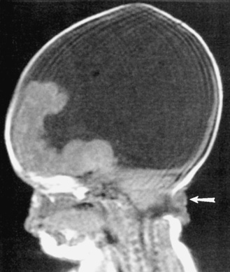
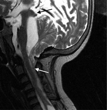
CHIARI II MALFORMATION
Definition
 the medulla is invariably elongated and kinked
Radiological features
CEREBRAL MALFORMATIONS
DISORDERS OF DORSAL INDUCTION
DEFINITION
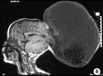
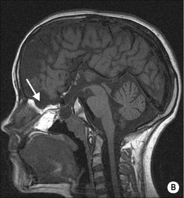
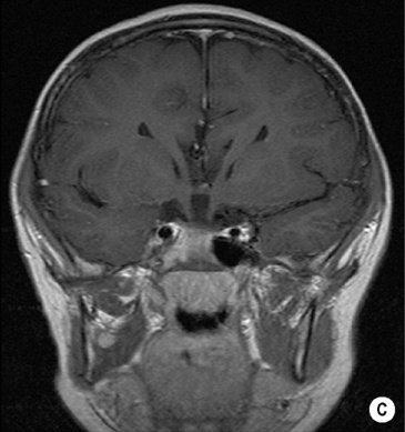
Cephalocele
 unlike a spinal myelomeningocele there is usually no skin defect
  they tend to occur in the occipital and frontal regions and may be pulsatile
DISORDERS OF VENTRAL INDUCTION
DEFINITION
Holoprosencephaly
 it is associated with chromosomal abnormalities, facial clefting and various teratogenic factors (including maternal diabetes)
MALFORMATIONS OF COMMISSURAL AND RELATED STRUCTURES
DEFINITION
PEARL
 thus a small or absent genu or body, with an intact splenium and rostrum, indicates secondary destruction rather than abnormal development
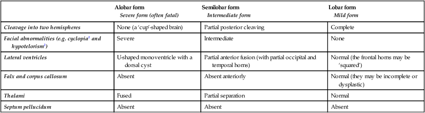
Alobar form
Severe form (often fatal)
Semilobar form
Intermediate form
Lobar form
Mild form
Cleavage into two hemispheres
None (a ‘cup’-shaped brain)
Partial posterior cleaving
Complete
Facial abnormalities (e.g. cyclopia1 and hypotelorism2)
Severe
Intermediate
None
Lateral ventricles
U-shaped monoventricle with a dorsal cyst
Partial anterior fusion (with partial occipital and temporal horns)
Normal (the frontal horns may be ‘squared’)
Falx and corpus callosum
Absent
Absent anteriorly
Normal (they may be incomplete or dysplastic)
Thalami
Fused
Partial separation
Normal
Septum pellucidum
Absent
Absent
Absent
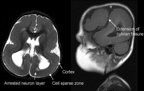
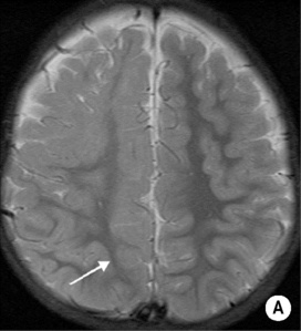
MALFORMATIONS OF NEURONAL MIGRATION AND CORTICAL ORGANIZATION
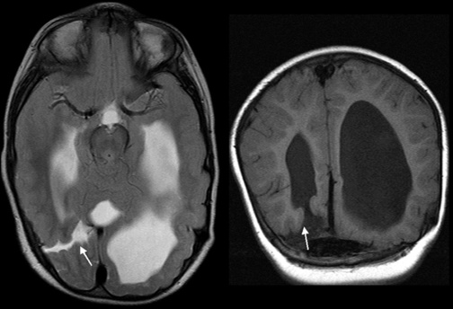
SCHIZENCEPHALY
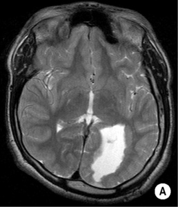
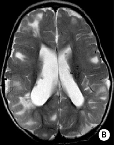
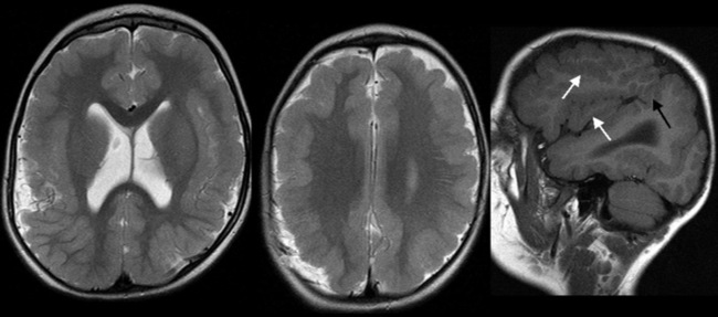
GREY MATTER HETEROTOPIAS
Band heterotopia or ‘double cortex’
 the overlying cortex is usually of normal thickness but has shallow sulci
  partial heterotopias predominantly affect the frontal lobes
Radiology Key
Fastest Radiology Insight Engine

 inborn errors of metabolism (e.g. glycolysation disorder)
 infantile neuroaxonal dystrophy
  pontocerebellar hypoplasia
  spinocerebellar atrophies
  Friedreich’s ataxia
 a normal-sized posterior fossa
 seizures
  hydrocephalus
 the cerebellar vermis is hypoplastic as well as rotated or aplastic
  the tentorium and venous confluence of the torcula are elevated
 the cerebellum and 4th ventricle are normal
 abnormal eye movements
  ataxia
 the midbrain is small
  the superior peduncles appear enlarged
 it is associated with other midline supratentorial anomalies (e.g. absence of the septum pellucidum and corpus callosum, as well as holoprosencephaly)

 an elongated medulla oblongata can be seen with a kink sometimes forming on its posterior surface
 basilar invagination (30%)
  hydrocephalus (25%)
  Klippel–Feil anomaly (10%)
 feeding problems
  dysphagia
 a small 4th ventricle (which is inferiorly displaced and elongated) and a small posterior fossa
  scalloping of the clivus
  flattening of the ventral pons and a low attachment of the tentorium
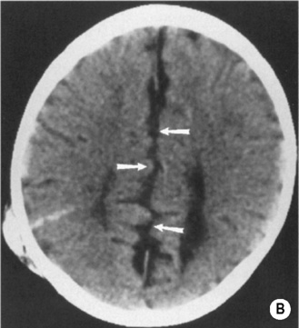
  the falx is partially absent or fenestrated with consequent interdigitation of the gyri across the midline
  the foramen magnum is enlarged and ‘shield-shaped’
 disorders of neuronal migration
  malformation of the corpus callosum
  a dorsal midline cyst
  absence of the septum pellucidum
  colpocephaly (occipital horn enlargement)
 an isolated 4th ventricle
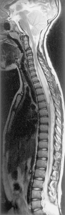
  hydro-syringomyelia
  compression of the craniocervical junction
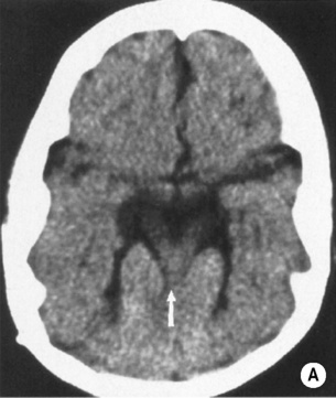
 there is no cerebral cortex present (unlike gross hydrocephalus)
 other intracranial malformations or hydrocephalus
  whether there is any ischaemia within the herniated neural tissue
 Dandy–Walker malformation
  interhemispheric lipoma
  abnormalities of neuronal migration and organization
  dysraphic anomalies
  encephaloceles
  septo-optic dysplasia
  ocular anomalies
  midline facial anomalies
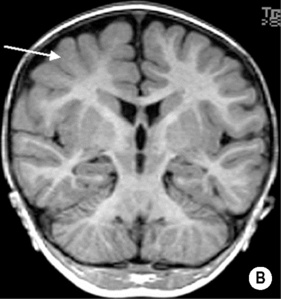
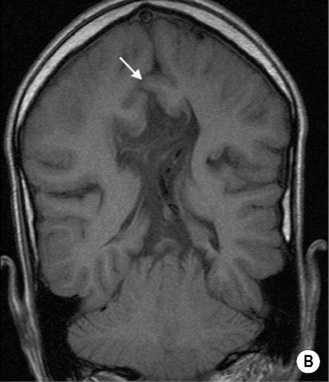
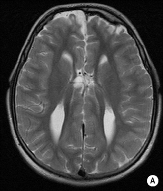
 vertically oriented sulci extend right down to the ventricle with no horizontally running cingulate sulcus
  small frontal horns (‘bull’s horn’ appearance) with colpocephaly (large occipital horns)
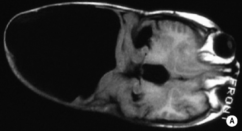
 it involves the complete cerebral mantle and connects the calvarium and the outer surface of the brain with the lateral ventricles
 it is also found within the parasagittal, frontal or occipital sites (with mild clinical manifestations)
 spasticity
  severe mental and psychomotor developmental delay (with bilateral clefts)
 it is associated with subependymal heterotopias (within the contralateral hemisphere) and subependymal or parenchymal calcification
 it is associated with agenesis or hypoplasia of the corpus callosum and septum pellucidum
 heterotopias are prominent and there is often a delay in myelination
  it may be seen in congenital muscular dystrophies
 T1WI + Gad: there is no enhancement
 they are also more heterogeneous and may enhance
 it may coexist with schizencephaly, microcephaly, polymicrogyria, dysgenesis of the corpus callosum, or absence of the septum pellucidum
 the extent varies from small, isolated, unilateral areas to larger areas of bilateral disease
 there may calcification or abnormal venous drainage
 other associations include neurofibromatosis type 1 (NF-1) and tuberous sclerosis
 the affected hemisphere is usually (but not always) enlarged with diffuse cortical thickening, white matter signal abnormality and possibly calcification