Presentation and Presenting Images
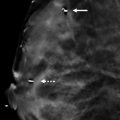
( ▶ Fig. 83.1, ▶ Fig. 83.2, ▶ Fig. 83.3)
A 78-year-old male who felt a lump in his right breast while showering presents for diagnostic mammographic evaluation.
83.2 Key Images
( ▶ Fig. 83.4, ▶ Fig. 83.5, ▶ Fig. 83.6, ▶ Fig. 83.7, ▶ Fig. 83.8)
83.2.1 Breast Tissue Density
There are scattered areas of fibroglandular density.
83.2.2 Imaging Findings
The imaging of the left breast is normal (not shown). The palpable finding in the right breast is denoted by the triangular marker (arrow in ▶ Fig. 83.4 and ▶ Fig. 83.5). The right breast demonstrates a high-density mass with indistinct margins and heterogeneous calcifications (circle) at the 7 o’clock position in the middle depth 2 cm from the nipple ( ▶ Fig. 83.4, ▶ Fig. 83.5, and ▶ Fig. 83.6). The right digital breast tomosynthesis (DBT) confirms with the mammographic findings and demonstrates that the mass has indistinct margins and corresponds to the palpable abnormality felt by the patient. The triangular marker localizes to the inferior aspect of the right breast ( ▶ Fig. 83.7 and ▶ Fig. 83.8).
83.3 Diagnostic Images
( ▶ Fig. 83.9)
83.3.1 Imaging Findings
Sonography demonstrates a hypoechoic mass with angular and indistinct margins ( ▶ Fig. 83.9). It corresponds to the mass seen mammographically.
83.4 BI-RADS Classification and Action
Category 5: Highly suggestive of malignancy
83.5 Differential Diagnosis
Breast cancer: Although rare, breast cancer does occur in men. This palpable mass is highly suspicious for malignancy.
Gynecomastia: Gynecomastia is usually eccentric to the nipple and retroareolar in location.
Simple cyst: The mass is hypoechoic and does not meet strict criteria for a cyst at ultrasound. Simple cysts are round or oval circumscribed masses with an imperceptible wall and posterior acoustic enhancement.
83.6 Essential Facts
Gynecomastia is described as hyperplasia of the ductal and stromal elements of the male breast. It typically presents as a soft, mobile, painful mass.
Male breast cancer is significantly less common than gynecomastia. It accounts for 1% of all breast cancers and 0.17% of all cancers in men.
Male breast cancer typically presents as a hard, fixed, painless mass.
Most male breast cancers are either in situ or invasive ductal carcinomas, because breast lobules are rare in males.
83.7 Management and Digital Breast Tomosynthesis Principles
DBT can be performed on male breasts.
As the parts of the breast tissue are viewed separately in slices with DBT, suspicious lesions may be quickly identified and accurately characterized.
The conventional mammographic and DBT images can be acquired with one compression.
The addition of DBT to conventional mammography significantly improves the diagnostic accuracy of conventional mammography.
83.8 Further Reading
[1] Appelbaum AH, Evans GFF, Levy KR, Amirkhan RH, Schumpert TD. Mammographic appearances of male breast disease. Radiographics. 1999; 19(3): 559‐568 PubMed
[2] Stavros AT, Rapp CL, Parker SH. Breast Ultrasound. Philadelphia, PA: Lippincott, Williams, & Wilkins; 2004.

Fig. 83.1 Right craniocaudal (RCC) mammogram.
Stay updated, free articles. Join our Telegram channel

Full access? Get Clinical Tree


