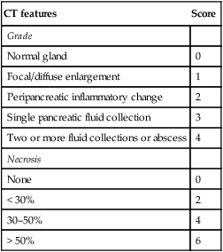
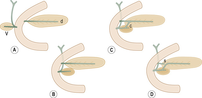
• The pancreas develops in two parts from the endoderm of the primitive duodenum • Dorsal part: this is the first part to appear, initially appearing as a diverticulum from the dorsal wall of the duodenum • Ventral part: this develops more caudally and initially appears as a diverticulum from the developing bile duct – Before this occurs the dorsal duct (the duct of Santorini) opens into the duodenum proximal to the major papilla (the ampulla of Vater) – The ventral duct (the duct of Wirsung) opens into the major papilla with the CBD • British Society of Gastroenterology Guidelines: immediate CT is not indicated as the full extent of necrosis is only evident after 4 days (therefore the initial extent of necrosis may be underestimated) • Mild acute pancreatitis (70–80%) • Necrotizing acute pancreatitis (a hallmark of severe acute pancreatitis) • Interstitial edematous pancreatitis (IEP) • Acute necrotizing pancreatitis (sterile or infected) • Pancreatic and peripancreatic collections (sterile or infected) – Acute peripancreatic fluid collections (APFC):presents within 4 weeks and usually resorbed spontaneously without infection – Pseudocyst: after 4 weeks, an APFC may transition to a pseudocyst with a well-defined enhancing fibrous wall (containing no non-liquefied components) Balthazar CT severity index (CTSI)
Pancreas
CONGENITAL ABNORMALITIES
EMBRYOLOGY
 it forms the neck, body and tail of the gland and part of the head
 it forms the remaining part of the head and uncinate process
 The duodenum undergoes partial rotation and the 2 parts approximate each other and fuse
PANCREATITIS
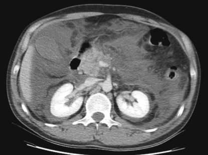
ACUTE PANCREATITIS
Radiological features
 the contrast medium may also exacerbate any renal impairment
 An immediate CT should only be performed if the extent of necrosis dictates the management or if the diagnosis is unclear
 A follow-up CT is only required if there is a failure to improve or a clinical deterioration
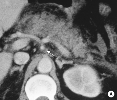
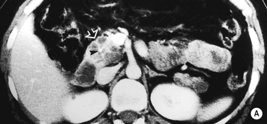
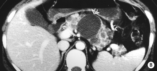
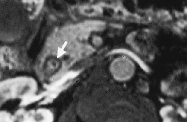
 A normal or enlarged gland of uniform enhancement (± peripancreatic fat stranding and thickening of the fascial planes)
  cuffs of fluid may be seen around the adjacent vessels
Pearls
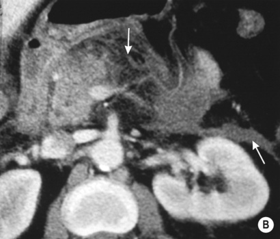
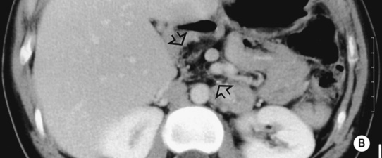
 Parenchymal necrosis alone – non-enhancing pancreatic areas
 Peripancreatic necrosis alone – commonly within retroperitoneum/lesser sac
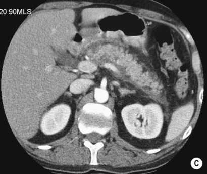
 no non-liquefied components
  usually adjacent to the pancreas
  no discernable wall
 these rarely become infected
ACUTE PANCREATITIS
CT features
Score
Grade
Normal gland
0
Focal/diffuse enlargement
1
Peripancreatic inflammatory change
2
Single pancreatic fluid collection
3
Two or more fluid collections or abscess
4
Necrosis
None
0
< 30%
2
30–50%
4
> 50%
6

 it is the commonest congenital pancreatic anomaly
  it may result in functional stenosis and pancreatitis and there is an increased incidence of pancreatic malignancies
 this is the 2nd most common congenital anomaly
 oesophageal atresia
  tracheo-oesophageal fistula
  Down’s syndrome
 85% of patients have severe exocrine pancreatic insufficiency and steatorrhoea
 dystrophic calcification
  pancreatic cysts
 serous cystic pancreatic neoplasms and pancreatic islet cell tumours may also occur
 hepatic cysts may also occur and pancreatic cysts are seen in 10% of patients
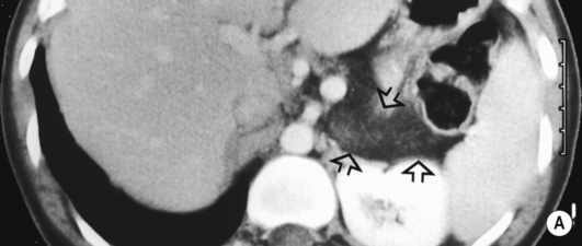
 it may be caused by the reflux of bile and pancreatic enzymes into the pancreatic parenchyma
 idiopathic (10–30% – possibly related to congenital duct anomalies such as pancreas divisum)
  alcohol (20–25%)
  trauma
  surgery
  metabolic (hyperlipidaemia and hypercalcaemia)
  viral infection (mumps, cytomegalovirus and AIDS)
  drugs (steroids and thiazide diuretics)
 nausea and vomiting
  raised serum amylase
  signs of haemorrhagic pancreatitis:
 a gasless abdomen
  ascites
  intrapancreatic gas bubbles
 ill-defined pancreatic margins
  peripancreatic fluid
  hepatic steatosis (if there is an associated high alcohol intake)
 if this involves > 30% of the gland the mortality rate approaches 30%
 it is a major determinant of morbidity and mortality


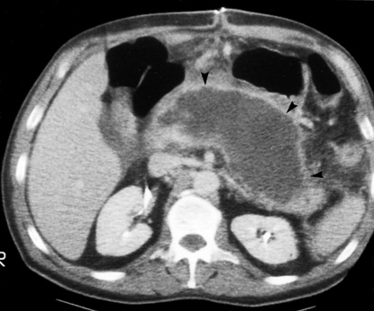
 liquefaction within 2–6 weeks
  any collection replacing pancreatic tissue within 4 weeks is an ANC and not an APFC/pseudocyst


 percutaneous drainage and supportive measures as required