Parapharyngeal Space
The parapharyngeal space is a triangular fat-filled space that extends from the skull base superiorly to the level of the hyoid bone, and is subdivided into the prestyloid (parapharyngeal [PPS]) space and the post-styloid (carotid space [CS]). In addition to fat, the major contents of interest in the PPS are minor salivary glands. Medially is the pharynx, posteromedially the retropharyngeal space, posterolaterally the CS, and laterally the masticator space. PPS displacement medially occurs by masses in the masticator space, the PPS may be displaced anteriorly by deep lobe parotid masses or masses in the CS and lateral displacement is caused by pharyngeal mucosal space lesions, particularly tonsil pathology.
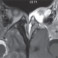
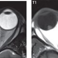
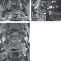
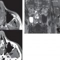
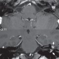
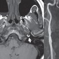
Salivary gland tumors (arising from the minor salivary glands) are the most common primary lesions in the PPS, with most being benign mixed tumors. Neurogenic tumors (usually vagal schwannomas) ( Fig. 2.97 ) and glomus vagale paragangliomas are the most common lesions to arise in the CS. Second branchial cleft cysts typically produce medial displacement of the CS, as well as lateral or posterior displacement of the sternocleidomastoid muscle and anterior displacement of the submandibular gland. One must be careful making this diagnosis in an adult as a level II necrotic node may mimic a branchial cleft cyst.

Related posts:
Stay updated, free articles. Join our Telegram channel

Full access? Get Clinical Tree


