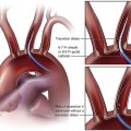
In experienced hands, primary surgical excision is curative in 95% of cases, but only 64% of patients with secondary surgery show success.1 Recently this outcome has been improved with the use of iPTH assays.2 When the primary surgery fails, reoperative parathyroidectomy is often difficult and unsuccessful because of scarring and distortion of the tissue planes without localization of the site of excess parathyroid hormone secretion by selective venous sampling (SVS). In a study of 228 consecutive patients with persistent/recurrent hyperparathyroidism, the single most common site of missed adenoma glands was in the tracheoesophageal groove in the superior compartment of the posterior mediastinum (27%). In this position, the glands may be adherent to the recurrent laryngeal nerve. Other ectopic sites for parathyroid adenomas in this group of patients were thymus (17%), intrathyroidal (10%), undescended glands (8.6%), carotid sheath (3.6%), and retroesophageal space (3.2%). The most sensitive and specific noninvasive imaging test was the 99mTc-sestamibi subtraction scan, with 67% true-positive and no false-positive results. The rate of true-positive results for ultrasonography, CT, MRI, and technetium thallium scans was approximately 50%.3 The accuracy of SVS for localization of the residual adenomas ranges between 75% and 90%4,5 and is related to the skill and persistence of the operator. It is possible to combine ultrasonography and needle aspiration for parathyroid hormone assay to identify lymph nodes that are the major cause of false-positive scans with ultrasonography, CT, and MRI.6 An ice bucket for the blood samples is required because all samples (usually 20-25) should be placed immediately on ice and transported to the laboratory with a vein diagram (Fig. 110-1) as soon as possible after the procedure.
Parathyroid Venous Sampling
Equipment
Catheter
Other Equipment
![]()
Stay updated, free articles. Join our Telegram channel

Full access? Get Clinical Tree


Parathyroid Venous Sampling


