Fig. 3.1
Patient positioned in low lithotomy position with a split leg attachment. The arms are positioned anatomically alongside the body and carefully secured. The vacuum mattress is inflated for cushioning. From John H. et al. Atlas of Robotic Prostatectomy. 2013;19–26. With permission from Springer Science + Business Media, LLC
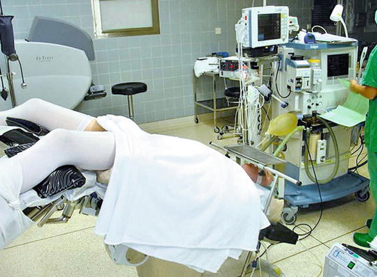
Fig. 3.2
Steep Trendelenburg positioning. From John H. et al. Atlas of Robotic Prostatectomy. 2013;19–26. With permission from Springer Science + Business Media, LLC
Securing the patient to the table can be safely performed in various ways. The patient’s chest may be strapped to the table either transversely or in a crossing “X” fashion. Alternatively, an inflatable bean bag can be secured around the patient including above the shoulders to prevent movement during positioning (Fig. 3.1). As the patient’s arms are tucked at the patient’s side, it is important that pulse oximetry, blood pressure cuff placement, and secure intravenous access be established prior to final positioning.
Trendelenburg positioning is necessary to ensure the bowel falls gravitationally out of the pelvis during the procedure (Fig. 3.2). Trendelenburg positioning is generally the extreme of the table, between 30 and 45°. Less Trendelenburg may be utilized depending on a patient’s ability to tolerate the positioning from a cardiopulmonary standpoint. In general, maximal Trendelenburg of a table will allow for correct positioning; however measurement of angulation using a nautical inclinometer was been employed [2].
Trendelenburg position is associated with numerous physiological effects about which the surgeon and anesthesiologist should be aware. These effects may impair cerebrovascular, respiratory, and hemodynamic function and may be exacerbated by pneumoperitoneum [4]. Cerebrovascular effects of Trendelenburg include increased intracranial pressure, which may be dangerous in patients with cerebral ischema or cerebrovascular disorders. However, overall there have been conflicting findings regarding the impact of Trendelenburg on cerebral oxygenation. Respiratory effects with Trendelenburg and pneumoperitoneum include mechanical pressure on the diaphragm, reducing functional reserve capacity and compliance, and predisposing patients to atelectasis. Difficulty ventilating may necessitate relieving some pneumoperitoneum or decreasing bed tilt. Hypercarbia may occur from CO2 insufflation and necessitate adjustments in minute and tidal volumes and/or adjustments of insufflation pressure by the surgeon. Effects on dead-space ventilation and oxygenation are thought to be small and are not typically an obstacle to positioning [3].
Hemodynamic effects occur with Trendelenburg in concert with pneumoperitoneum as well. Mean arterial pressure and systemic vascular resistance increase when pneumoperitoneum is initiated; adding Trendelenburg may return systemic vascular resistance to baseline but not change mean arterial pressure [6]. While central venous pressure, mean pulmonary arterial pressure, and pulmonary capillary wedge pressure do not change with insufflation, adding steep positioning may cause a significant increase in these values [6]. Heart strain may increase with Trendelenburg positioning as well. In general, patients with normal cardiac function tolerate these perturbations; however, those with compromised heart function may be at risk for heart failure from preload increase with positioning [6]. Finally, bradycardia may be induced as a vagal response to pneumoperitoneum; this generally subsides with relief of intraabdominal pressure. It is prudent for patients with a history of significant cardiopulmonary disease to consult with cardiology and/or pulmonology prior to surgery to address the safety of Trendelenburg positioning.
Additional risks of steep Trendelenburg include facial, pharyngeal, and laryngeal edema, associated with venous congestion, facial swelling, and increased intracranial pressure. Restricting fluids and minimizing operative time may help to minimize perioperative edema.
Patient movement during Trendelenburg positioning should be avoided, in particular to avoid instability of the head and neck. Obese patients may be at greater risk for migration on the table. Generally, however, the patient remains stable after docking of the robot. Meticulous attention in securing the patient prior to bed tilt is essential to minimize patient movement.
A potential complication with steep Trendelenburg positioning is compartment syndrome and/or rhabdomyolysis, which may be life threatening. Longer operative times and high body mass index may be risk factors for this complication [12]. In patients with longer case duration, particularly for obese patients, there should be a higher index of suspicion for rhabdomyolysis, and surveillance serum creatine kinase measurements and aggressive postoperative hydration should be considered [12]. Gluteal and calf fasciotomies have been required for compartment syndrome in isolated reports [5, 8].
Patients with a history of glaucoma are at theoretical risk for exacerbation of their disease with Trendelenburg positioning. Studies of intraocular pressure have demonstrated that surgical duration and end-tidal CO2 are significant predictions of intraocular pressure increase in the Trendelenburg position [1]. However, the precise risks of positioning in these patients are unclear. While it is thought that RALRP is safe in patients with controlled glaucoma, patients with severe or uncontrolled disease should consult with ophthalmology prior to surgery. Additionally, there has been one reported case of ischemic optic neuropathy during RALRP, with prolonged positioning, substantial intravenous fluids, and substantial blood loss [10]. Ultimately, avoidance of protracted positioning is physiologically advantageous to the patient and an expeditious procedure is likely to minimize risk of ocular and other complications.
Positioning for Radical Retropubic Prostatectomy
For standard positioning, the patient is placed in the supine position. Traditionally the patient is placed with his hip on the break of the table, and the table is flexed to increase the distance between pubis and umbilicus. However, flexion of the table is not universally performed. If the table is flexed (i.e., hyperlordotic positioning), the kidney rest may be elevated to increase puboumbilical distance to improve exposure. However, while this is the classical approach to positioning, there have been reports of femoral and sciatic nerve injuries, and rhabdomyolysis with acute renal failure in this position [9]. Obese patients may be at increased risk for rhabdomyolysis and nerve palsy syndromes with RRP, thus require extra care with positioning. Another concern with hyperlordotic positioning is that with severe lumbar stenosis, extension of the spine may compress neural elements and potentially cause spinal cord trauma [9]. Thus the authors generally recommend supine positioning without table flexion.
Related posts:
 Challenging Cases in Robotic Radical Prostatectomy
 Postoperative Management and Preoperative Considerations: Urinary Incontinence and Anastomotic Stricture
 Pelvic Lymph Node Dissection for Prostate Cancer
 The Transperitoneal Robotic-Assisted Radical Prostatectomy
 Surgical Anatomy
 Open Radical Retropubic Prostatectomy
Stay updated, free articles. Join our Telegram channel

Full access? Get Clinical Tree


