Hypoplastic acetabular roof with later development of increasing sclerosis. Posterosuperior dislocation of the femoral head ± flattening of the femoral head.
Ultrasound (US) is the screening modality of choice before ossification excludes visualization of the hip structures (below the age of 6 mo).
Secondary hip dysplasia
Increased acetabular angle. Shallow acetabulum. Coxa valga, ± femoral head dislocation and flattening.
Generally occurs in neuromuscular diseases with spasticity of the lower extremities. May develop in a previously normal hip.
Arthrogryposis
Very thin diaphyses, osteopenia, dislocation and subluxation of large joints.
Joint contractures and decreased muscle bulk.
Ehlers-Danlos syndrome types III and VII
Hip dislocation with an otherwise normal appearing skeleton.
Calcified round areas of subcutaneous fat necrosis.
Accessory carpal ossification centers, double calcaneal ossification center. Dislocation of large joints.
Flat facies. Clinical DD: Marfan syndrome, arthrogryposis, and Ehlers-Danlos syndrome.
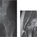
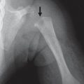
Fig. 5.20a–c Developmental dysplasia of the left hip with dislocation and delayed ossification of the proximal femoral epiphysis (a). (b) US shows dislocation. (c) Normal side for comparison.Fig. 5.21 Developmental dysplasia of the left hip with a shallow acetabulum.
Lateral and Medial Spurs of the Acetabulum (Trident Acetabulum) Acetabular Protrusio
Spurs are seen in skeletal dysplasias when the base of the iliac bone is small and the entire iliac bone is decreased in size. The spur is visible in neonates and young infants and disappears as the skeleton matures.
Only gold members can continue reading. Log In or Register to continue