Abstract
Pentalogy of Cantrell is a rare syndrome consisting of a spectrum of five structural ventral midline defects, with the combination of an omphalocele and ectopia cordis being the hallmark of the disorder. Although the pathogenesis of this syndrome is not well understood, early embryologic developmental failure of a segment of the lateral mesoderm is a prevailing theory. Prenatal diagnosis relies on sonographic and echocardiographic findings and can be suspected as early as 12–13 weeks’ gestation. A responsible gene has not yet been identified; therefore no genetic testing is available to provide a definitive prenatal diagnosis. The overall survival rate is poor and is directly correlated to the type and complexity of the thoracoabdominal defects and any associated intracardiac abnormalities. A multidisciplinary team approach is essential for accurate diagnosis as well as prenatal and postnatal care.
Keywords
abdominal wall defect, diaphragmatic hernia, ectopia cordis, omphalocele, pentalogy of Cantrell
Introduction
First described in 1958, pentalogy of Cantrell is a rare syndrome consisting of five anomalies including: (1) a midline supraumbilical abdominal wall defect, (2) a defect in the diaphragmatic pericardium, (3) a defect in the lower sternum, (4) a defect in the anterior diaphragm, and (5) various intracardiac anomalies. In the most severe cases, the heart herniates through the diaphragmatic defect, resulting in ectopia cordis. The association of an omphalocele with ectopia cordis is the hallmark of this syndrome. Multiple variants have been reported, and only a few patients display the complete spectrum of disease. The mortality rate is high; however, survival and prognosis ultimately depend on the type and complexity of the associated defects.
Disorder
Definition
The full pentad of the syndrome of pentalogy of Cantrell is rare and consists of the following five defects:
- 1.
deficiency of the anterior diaphragm,
- 2.
defect of the lower sternum,
- 3.
midline supraumbilical abdominal wall defect,
- 4.
defect in the diaphragmatic pericardium,
- 5.
congenital intracardiac abnormality.
Class 1: definite diagnosis (all five defects present).
Class 2: probable diagnosis (four defects present, including ventral wall abnormalities and cardiac defects).
Class 3: incomplete expression (various combinations of defects present, including a sternal abnormality).
Prevalence and Epidemiology
The prevalence of pentalogy of Cantrell has been reported to range from 1 : 65,000 to 5.5 : 1 million live births. This wide range is secondary to the variation in classification of the syndrome and whether estimates are made using the strictest criteria (class 1) versus more liberal criteria (class 3). A 2 : 1 male predominance has been reported in some series.
Etiology, Pathophysiology, and Embryology
The etiology and pathogenesis for this syndrome is not well understood and is thought to be of a heterogeneous origin. On an embryologic basis, Cantrell et al. proposed that the association of defects is mesodermal in origin, occurring in the third week of embryonic life. Abnormal migration of splanchnic and somatic mesoderm with premature breakage of the vitelline sac results in a midline defect. Abnormal formation of the transverse septum of the diaphragm occurs because of abnormal migration of myoblasts; premature atrophy of the cardinal vein leads to associated pericardial defects.
Most reported cases of pentalogy of Cantrell are thought to be sporadic in occurrence; however, mutations in genes located on the X chromosome have been associated with ventral midline disruptions. An X-linked dominant inheritance pattern has been observed in some families. In such cases, the ventral midline is viewed as a developmental field, and embryologic disruptions in single genes involved in the development of this field are responsible for the wide range of midline defects. It is unclear whether pentalogy of Cantrell represents a separate entity as opposed to an extreme end of the spectrum of ventral midline disorders.
A responsible gene for this disorder has not yet been identified; however, a recent case report demonstrated a novel maternally-in herited microduplication of chromosome 15q21.3 on postnatal chromosomal microarray of a newborn with a prenatal diagnosis of pentalogy. This region includes the gene ALDH1A2 , which encodes for retinaldehyde dehydrogenase type 2, an enzyme with a critical role in normal cardiac and diaphragm development, thereby demonstrating a biologically plausible association with this disorder.
Manifestations of Disease
Clinical Presentation
There is tremendous variability in the number and type of defects observed in pentalogy of Cantrell. Common findings include :
- •
abdominal wall defects (giant omphalocele, diastasis of the rectus muscles),
- •
anterior diaphragmatic hernia,
- •
anterior sternal defect with the heart protruding outside of the thoracic cavity,
- •
displacement of the heart through a diaphragmatic defect (thoracoabdominal ectopia cordis),
- •
cardiac malformations (ventricular septal defect, atrial septal defect, tetralogy of Fallot, ventricular diverticula, ventricular aneurysm).
- •
central nervous system malformations (neural tube defects, encephalocele, hydrocephalus),
- •
craniofacial defects (cleft lip and/or palate, craniorachischisis, exencephaly),
- •
limb defects (club foot, absence of tibia or radius, hypodactyly),
- •
abdominal organ defects (intestinal malrotation, polysplenia, gallbladder agenesis).
Imaging Technique and Findings
Ultrasound.
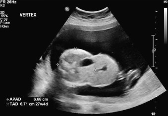
Sonographic diagnosis of pentalogy of Cantrell has been reported as early as 10–12 weeks’ gestation. Identification of an omphalocele and an ectopia cordis should raise suspicion for the diagnosis of pentalogy of Cantrell ( Figs. 136.1–136.3 ). The sonographic appearance of ectopia cordis is variable but, in its most severe form, can be identified as the fetal heart protruding through a wide sternal cleft outside of the thoracic cavity. In cases where ectopia cordis is not easily identifiable on ultrasound (US), the presence of a transient pericardial or pleural effusion in the setting of an omphalocele may be a marker for a coexisting diaphragmatic or pericardial defect. Increased nuchal translucency and cystic hygroma have also been associated with this syndrome and may serve as additional first-trimester markers.


