Percutaneous Abscess Drainage Within the Abdomen and Pelvis


Percutaneous Abscess Drainage Within the Abdomen and Pelvis


Justin J. Campbell and Debra A. Gervais


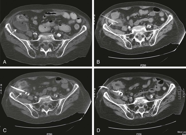
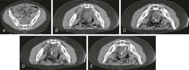
Since its introduction in the early 1980s, percutaneous abscess drainage (PAD) has become the primary drainage procedure for most infected abdominal fluid collections. Numerous large clinical series have demonstrated the efficacy and safety of this procedure.16 Image-guided drainage allows rapid, minimally invasive drainage of collections that had previously required surgical drainage and necessitated a trip to the operating room, general anesthesia, and was therefore associated with higher morbidity. Benefits of PAD in most cases include avoiding general anesthesia, laparotomy, and prolonged postoperative hospitalization, with a resultant reduction in morbidity, mortality, and hospital costs.


Indications


As PAD has become more widely used, indications have continued to expand. Currently, the vast majority of infected intraabdominal and pelvic fluid collections are considered amenable to percutaneous drainage. Prerequisites for PAD are presence of a fluid collection and one or more of the following: suspicion the collection is infected, need for fluid analysis, or suspicion the collection is producing symptoms that warrant drainage.7 PAD may be performed in essentially every organ of the abdomen and pelvis. Liver abscesses, pancreatic abscesses, renal and perirenal abscesses, enteric abscesses, anastomotic abscesses, subphrenic abscesses, splenic abscesses, and pelvic abscesses, as well as subcutaneous abscesses and abscesses within musculature (e.g., psoas muscle) are examples of abscesses that have been successfully drained with percutaneous techniques. The goal of treatment can be complete cure of an infected fluid collection, or PAD may be used as a temporizing measure before definitive surgical treatment. For example, patients with diverticulitis and abscess formation would require, in addition to surgical drainage, a colostomy and subsequent takedown of the colostomy if operated on in the acute period. With PAD, the abscess and any existing or potential acute sepsis are treated first, allowing time for the acute inflammation to subside and ensuring a cleansed bowel at the time of resection so a primary anastomosis can be performed without colostomy. In other instances, PAD can serve as a palliative procedure for patients who are not candidates for curative surgical treatment.


Contraindications


Contraindications to PAD are relative and should be weighed carefully against the suitable surgical alternative and the alternative of no procedure. Relative contraindications include coagulopathy that cannot be adequately corrected, phlegmons or collections containing a large amount of debris, hemodynamic instability such that the procedure could not be completed, inability of the patient to be positioned or cooperate adequately for the procedure, known adverse reaction to contrast material when administration is deemed necessary for success of the procedure, lack of a safe access route for drainage of a collection, and severely compromised cardiopulmonary status in patients undergoing procedures in which there is risk of further cardiopulmonary compromise inherent in the procedure.7 In reality, almost all these factors can be mitigated to some degree to allow abscess drainage. For example, most coagulopathies can be corrected to within an acceptable range of risk. In truth, the only absolute contraindication would be absence of a safe percutaneous route to the abscess. However, this is rare with the many options available to the interventional radiologist, such as angling of the computed tomography (CT) gantry, using an angled ultrasound approach, imaging after repositioning the patient, or instilling saline to displace structures at risk. The risks and benefits of PAD must be carefully evaluated in each patient. Decisions regarding whether to pursue abscess drainage in critically ill patients are best made in concert with the surgical and medical teams caring for the patient.


Additionally, imaging facilities should have policies and procedures in place to attempt to identify any pregnant patients before exposing them to ionizing radiation. Pregnant patients undergoing procedures requiring use of ionizing radiation are counseled regarding the risk of radiation exposure to the fetus and the clinical benefit of the procedure. Every attempt should be made to avoid exposing the fetus to radiation in accordance with ALARA (as low as reasonably achievable) principles. This can be accomplished by performing the procedure under ultrasound guidance alone when possible. If CT is required for adequate drainage, exposure factors are minimized to those required to demonstrate the collection and differentiate it from adjacent structures. Likewise, if fluoroscopy is needed to guide wire manipulations, diminished exposure factors and frame rates are advisable.


Equipment


The primary tool necessary for drainage of an abscess is a drainage catheter. In the early days of PAD, angiography catheters and catheters used by surgeons were the tools available for PAD, but catheters specifically designed for PAD are now available. The majority of catheters are constructed from a proprietary polyurethane material modified for kink resistance. These catheters are designed to be placed by either the Seldinger or trocar method. Standard catheters consist of a pointed metal trocar loaded coaxially through a metal stiffener into the flexible polyurethane catheter. Once the tip is placed in the collection, the catheter is fed off the trocar and stiffener into the collection. The trocar and stiffener are then removed. Alternatively, the catheter can be placed by the Seldinger method by removing the trocar and placing the catheter and stiffener over a wire. After dilating the tract, the catheter is fed off the stiffener once it has passed through any tissues likely to prevent the catheter from easily following the wire, such as fascial planes and muscle. Catheters are available in sizes ranging from 8F to 16F. Many have a hydrophilic coating to facilitate placement and deployment. Numerous large oblong-shaped holes at the catheter tip facilitate drainage. The majority of catheter tips have a string-locked pigtail configuration for retention, but other internal retention devices, such as balloons and mushroom types, are also available.


Whether using the trocar or Seldinger technique, needle access is generally performed before catheter placement. A variety of needles can be used. Superficial collections are generally amenable to access with spinal needles. Deeper collections can be sampled with 10- to 25-cm Chiba needles. Needle diameters range from 18 to 22 gauge. Fluid sampling is usually possible with 20-gauge needles, but if a viscous collection is suspected, larger-gauge needles can be used. The needle can then be used as a guide for the tandem trocar technique. If the Seldinger technique is performed, the access needle can be used to place the wire over which the drainage catheter will be placed. Several needles designed to accept a 0.038-inch wire can be used to facilitate this technique. Ring sheathed needles (Cook Medical, Bloomington, Ind.) allow direct placement of an 0.038-inch wire, whereas the AccuStick system (MediTech/Boston Scientific, Watertown, Mass.) allows initial placement of an 0.018-inch wire, with upsizing to a 0.038-inch wire via use of a sheath.


When the Seldinger technique is used, the operator may need to redirect a working wire to a specific location for optimal catheter drainage. This is usually achieved by using a directional catheter to guide the wire to a specific target location under fluoroscopic guidance. A number of such catheters exist. We find that a 5.0 Kumpe catheter or cobra catheter is useful for this purpose. These catheters accept 0.038-inch wires. In general, directional catheters measuring 30 to 40 cm in length are preferable to the longer catheters used in vascular interventions, because the distance from the skin puncture to the desired catheter location is usually much shorter than this. A catheter that is too long can make torquing and redirecting the catheter more difficult.


When using the Seldinger technique, tract dilation is often necessary to allow placement of the catheter. This can be achieved with dilators, stiff devices with tapered ends that are placed over the working wire. During tract dilation, care must be taken to not kink the wire. After performing serial dilation, the catheter can then be placed over the wire. Alternatively, the catheter can be placed after dilation with the use of a Peel-Away introducer (Cook Medical). The advantage of the latter is that it provides added stiffness for passing the catheter through tracts that are especially resistant to the softer catheter. This will also require the tract to be dilated 1 to 2 French sizes larger than the nominal drainage catheter size.


Once the catheter has been placed within the collection and secured with the internal retention device, it is usually fixed to the skin to provide additional stability to prevent dislodgment of the catheter from the collection. This can be performed in a number of different ways. The catheter can be secured with a skin suture, which is then attached directly to the catheter. Alternatively, nonsuture fixation devices can be used. These devices are fixed to the skin with adhesive and lock to the catheter with a plastic locking device designed to accommodate specific catheter sizes. One example is the StatLock system (Venetec, San Diego, Calif.). A hybrid system can be used in which the catheter is sutured to an adhesive ostomy appliance (Hollister Inc., Libertyville, Ill.) that contains a small central opening through which the catheter passes. Finally, a drainage bag is attached to the catheter. A three-way stopcock can be placed between the catheter and bag to facilitate catheter flushing. At our institution, abdominal/pelvic drainage catheters are usually placed under gravity drainage, but some practitioners prefer using suction devices.


Technique


Anatomy and Approach


After establishing the presence of a drainable fluid collection, several issues regarding patient preparation must be addressed before performing PAD. First, adequate antibiotic therapy is initiated if there is clinical evidence of infection. In the majority of cases, the team caring for the patient has begun antibiotic therapy before consulting radiology for potential PAD. In the rare instance in which antibiotic therapy has not been initiated, broad-spectrum antibiotics are administered before drainage. Antimicrobial therapy is not withheld for fear of sterilizing cultures; because the interventional manipulation can seed the bloodstream with bacteria, the risk of sepsis is too high.8 Coagulation factors, including prothrombin time (PT), partial thromboplastin time (PTT), and platelet count, are checked before intervention. Any coagulopathy is then corrected to the degree possible before performing the procedure.


Except for the most superficial abscesses, most abscess drainage procedures cause some intraprocedural pain despite local analgesia, so intravenous (IV) sedation is almost always used. A benzodiazepine and narcotic combination works well. At our institution we use IV fentanyl citrate and midazolam hydrochloride (Versed). Typically, fentanyl (25 µg IV) and midazolam (0.25 to 0.5 mg IV) are administered at 5- to 10-minute intervals until adequate sedation is achieved. IV sedation is administered according to institutional guidelines, and interventional radiologists need to be familiar with their own institution’s guidelines.


When planning the drainage route, it is important to consider the most direct route that is free of intervening organs and vital structures such as bowel, solid viscera, vessels, and nerves. Furthermore, every attempt is made to avoid contaminating sterile areas. The catheter is generally placed in the most dependent position possible to facilitate drainage. For example, if the patient will be in the supine position after placing the catheter, a posterior or lateral approach may optimize drainage. Once the route for drainage has been selected, the patient can be placed in the most appropriate position for catheter placement, after which the skin is prepared and draped in the usual sterile fashion, and sedation is initiated.


Initial access to the abscess cavity is obtained under CT or ultrasound guidance. Fluoroscopy can be used in conjunction with either of these two modalities to confirm catheter position or assist with catheter positioning in difficult cases. The decision regarding which imaging modality to use is based on factors that include position of the collection, presence of overlying structures, visibility of the collection and adjacent structures with each modality, availability, and user preference. A limited number of image-guided drainage procedures are performed under magnetic resonance imaging (MRI) guidance, but such techniques are still in the early stages of development and not yet widely used. The decision of which modality to use should be made on a case-by-case basis.


Ultrasound has several advantages. First, it is a real-time imaging modality and for this reason is generally much faster than CT. It is also more widely available and less expensive than CT. The modality is portable, allowing for use at the bedside in critically ill patients not stable enough to travel to the radiology suite. Finally, ultrasound does not expose the patient to ionizing radiation. Unlike CT, however, intervening fat, gas, and bone significantly degrade visualization of collections. Abscesses deep within the abdomen or pelvis can be difficult to visualize. For the same reason, ultrasound can be difficult to use in obese patients with large amounts of subcutaneous fat. Similarly, collections in the retroperitoneum can be difficult to visualize. Limitations of ultrasound also include the need for significant operator experience.


CT is not as operator dependent as ultrasound, and collections deep to bone, gas, or fat are well visualized. Provided the patient’s weight does not exceed table limits and his or her size allows placement of the patient with a guiding needle into the gantry, CT-guided drainage is not as limited by obesity as ultrasound-guided drainage. Lastly, both final catheter position and the degree of evacuation of abscess contents are better visualized with CT than with ultrasound. Disadvantages of CT include exposure to ionizing radiation, increased time required for procedures, and lack of portability.


Both CT and ultrasound have limitations in their ability to distinguish solid tissue from drainable fluid collections. CT relies on attenuation measurements to distinguish tissues. The majority of fluid collections have low attenuation, but some drainable fluid collections can contain high-attenuation material such as iodinated contrast material or blood products. Likewise, some nondrainable tissues (e.g., necrotic tissue, certain neoplasms) can be very low in attenuation. Unlike CT, however, ultrasound has particular difficulty visualizing collections that contain gas because of limited sound transmission. Additionally, some hypoechoic through-transmitting tissues (e.g., lymphoma) can be mistaken for fluid. Regardless of which imaging modality is ultimately chosen to guide drainage, a combination of predrainage diagnostic imaging modalities can be used to characterize collections and the feasibility of drainage access before attempting PAD.


Technical Aspects


Method


The catheter may be placed by either the trocar or Seldinger technique. Both are viable options and offer advantages and disadvantages in different clinical settings. Although each technique has its proponents and detractors, no randomized trial has been conducted to compare them, and the choice of which to use comes down to operator preference, which itself is usually a function of experience. The trocar technique is generally performed in conjunction with a guiding needle (Fig. 127-1). As discussed earlier, the guiding needle is usually 18 to 22 gauge and variable in length, depending on the depth of the collection. The guiding needle is placed in the collection under CT guidance. Once needle placement within the collection has been confirmed with imaging, the needle acts as a guide for catheter placement. Before placing the catheter, a sample of fluid can be aspirated and sent for laboratory analysis. This initial aspiration can be helpful in charactering the fluid. If no fluid is aspirated, the collection may be very viscous and thus suggest the need for a larger catheter. Alternatively, if the fluid is not grossly purulent and the collection is not thought to be infected, a Gram stain can be performed if the decision of whether to place a catheter will be influenced by these results.


Once the decision to place the catheter has been made, a small nick is made in the skin adjacent to the entrance site of the guiding needle, and the underlying subcutaneous tissue is dissected bluntly. The catheter containing the hollow metal stiffener and inner trocar needle is then advanced into the cavity, adjacent to the guiding needle, to the appropriate predetermined depth. When the catheter tip is within the cavity, the catheter is fed off the trocar needle and metal stiffener. Syringe aspiration can then be performed to help confirm catheter position. Minimal fluid is aspirated at this time because decreasing the size of the target cavity makes subsequent repositioning technically more difficult if required. Catheter position is then confirmed by imaging before securing the device. The guiding needle is removed after satisfactory catheter position is confirmed. The advantage of the trocar method is the speed with which the catheter can be deployed. This can be very helpful when the patient is critically ill or lightly sedated. The main disadvantage of this technique is limited ability to reposition the catheter without removing and reinserting it if initial positioning is suboptimal.


With the Seldinger technique, initial access can be performed with a needle under ultrasound or CT guidance (Fig. 127-2). When needle position within the cavity is confirmed by imaging, a wire is placed through the needle into the cavity. The needle is then removed over the wire, and the tract is serially dilated to the appropriate size to allow placement of the catheter. Two different types of needles can be used for initial access. “One-stick” systems use a 21- to 22-gauge needle that accepts a 0.018-inch wire. A three-component device containing a sheath, dilator, and stiffener is placed over this wire. With removal of the stiffener and dilator, a 0.035- or 0.038-inch working wire can be placed through the sheath. Further dilation can then be performed to place the catheter over this wire into the collection. Alternatively, a larger needle that accepts a 0.035- or 0.038-inch wire, such as a Ring needle, can be placed. The Seldinger technique is helpful when the window to the collection is narrow. This technique offers the further advantage of guiding the catheter into optimal position with the use of directional catheters under fluoroscopic guidance. The disadvantage of this technique is primarily that it can be more time consuming than the trocar technique. This time consideration can be particularly important with critically ill patients. Additionally, serial dilation can be painful even in well-sedated patients.


Dec 23, 2015 | Posted by in INTERVENTIONAL RADIOLOGY | Comments Off on Percutaneous Abscess Drainage Within the Abdomen and Pelvis

Full access? Get Clinical Tree

Get Clinical Tree app for offline access