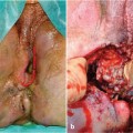
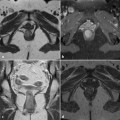
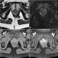
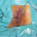
Fig. 1.1
The perineum
Dissecting the skin layer reveals the external anal sphincter and between it and the lateral border of the perineum the ischioanal fossa. This fossa has a prismatic shape, with its base directed towards the surface of the perineum and its apex meeting the obturator and anal fasciae. The lateral border is composed of the obturator internus muscle and its fascia. The pudendal vessels and nerves are located in a splitting of this latter fascia. The medial border is formed by the levator ani and the external anal sphincter. The ischioanal fossa has two extensions, an anterior one between the levator ani and perineal fascia, and a posterior one, between the levator ani and gluteus major [3].
1.2 The Urogenital Triangle
The musculotendinous layer of the urogenital triangle closes the anterior part of the pelvis. In males, it is crossed by the urethra and bulbo-urethral glands, and in females by the urethra, vagina, and vestibular glands. It is composed of several different layers: the deep transverse perineal muscle, between the bones of the ischium; the urethral sphincter surrounding the urethra; the ischiocavernosus muscle, extending from the inner surface of the ischial tuberosity to the pubic bones; and a bulbospongiosus muscle arising from the central tendinous point of the perineum. In males, it encircles the urethra and in females it covers the vestibular bulb and envelops the vagina [1].
1.3 The Anal Triangle
The most important structure of the anal triangle is of course the anal canal. Developmentally, it is a region of fusion between endodermal and ectodermal tubes, evident at the dentate line. The distal colon is derived from the hindgut and is thus made up of endodermal tissue. Before the 5th gestational week, the intestinal and urogenital tract flow in the cloaca. During the 6th gestational week, the two tracts separate. The anal canal is the terminal portion of the large intestine. It forms an angle with the lower part of the rectum, measures 2.5-4 cm, and is surrounded by the internal and external anal sphincters. A variable number of vertical folds, the rectal columns, lie 7–15 mm above the anal orifice and are separated from each other by rectal sinuses, which end in the anal valves.
Stay updated, free articles. Join our Telegram channel

Full access? Get Clinical Tree


