Mohini Rawat, DPT, MS, ECS, OCS, RMSK
Contents
- Upper Extremity Nerves
- Brachial Plexus and Thoracic Outlet
- Cervical Nerve Roots
- Vagus Nerve and Cervical Ganglion
- Greater Auricular Nerve
- Spinal Accessory Nerve
- Dorsal Scapular Nerve
- Facial Nerve
- Greater and Lesser Occipital Nerves
- Phrenic Nerve
- Subclavian Nerve
- Long Thoracic Nerve
- Cervical Nerve Roots
- Shoulder and Arm Region
- Elbow and Forearm Region
- Lateral Antebrachial Cutaneous Nerve
- Medial Antebrachial Cutaneous Nerve
- Posterior Antebrachial Cutaneous Nerve
- Ulnar Nerve
- Radial Nerve and Its Branches
- Median and Anterior Interosseous Nerves
- Wrist, Hand, and Digits
- Elbow and Forearm Region
- Brachial Plexus and Thoracic Outlet
- Lower Extremity Nerves
Brachial Plexus and Thoracic Outlet
- Probe/patient position: The patient’s head is rotated to the opposite side, and the probe is positioned on the sagittal oblique axis over the scalene anterior, perpendicular to the supraclavicular vessels, to visualize the cervical nerve roots in the short axis (SX) from C5-C8. Tracing the nerve roots proximally, C5 and C6 can be seen emerging between the anterior and posterior tubercles (Figure 8-1). The C7 nerve root lacks an anterior tubercle (Figure 8-2). Moving a few millimeters distally, the trunk level can be visualized in the long axis (LX) through the acoustic window of the scalene middle (Figures 8-3 through 8-5). Further distally, at the level of lateral third of the supraclavicular fossa and between the clavicle and first rib in the sagittal oblique view, 6 divisions of the brachial plexus appear as a cluster of hypoechoic fascicles between the omohyoid muscle and the subclavian artery and vein (Figure 8-6). The cord level of the brachial plexus is best visualized in the subpectoralis minor space, where the probe is placed in the sagittal oblique orientation over the pectoralis minor muscle and the axillary artery is identified (Figure 8-7). The posterior, medial, and lateral cords are identified around the axillary artery as hyperechoic structures.1–4
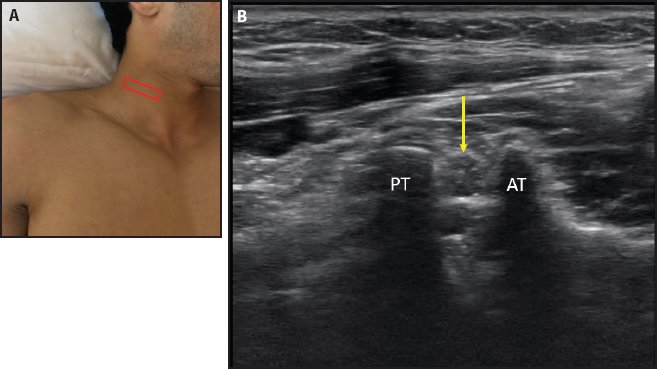
Figure 8-1. C5 nerve root. (A) Probe placement. (B) SX view of the C5 nerve root (yellow arrow) emerging between the anterior tubercle (AT) and posterior tubercle (PT).
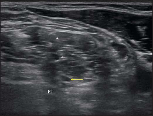
Figure 8-2. C7 nerve root (yellow arrow), posterior tubercle (PT), and overlying C5 and C6 nerve roots (white arrows).
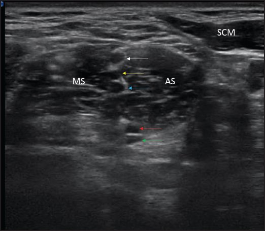
Figure 8-3. SX view of the C5 (white arrow), C6 (yellow arrow), C7 (blue arrow), C8 (red arrow), and T1 (green arrow) nerve roots emerging between the anterior scalene (AS) and middle scalene (MS) muscles. Also shown is the overlying sternocleidomastoid muscle (SCM).
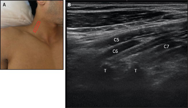
Figure 8-4. LX view of the C5, C6, and C7 nerve roots. (A) Probe placement. (B) LX view of the nerve roots as they emerge between the anterior and posterior tubercles (T).
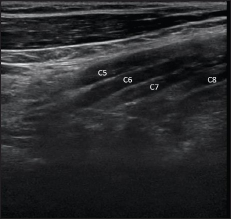
Figure 8-5. LX view, slightly distal to the view in Figure 8-4, showing C5, C6, C7, and C8 nerve roots.
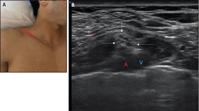
Figure 8-6. Six divisions of the brachial plexus. (A) Probe placement. (B) SX view at the division level (white arrows). The brachial plexus appears as a cluster of hypoechoic fascicles between the omohyoid muscle (red arrow), the subclavian artery (red A), and vein (blue V).
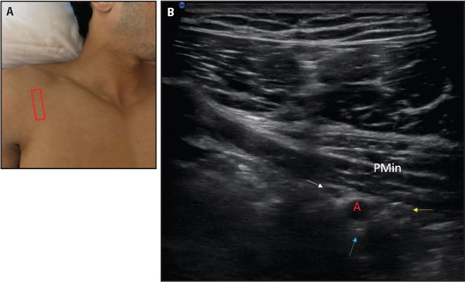
Figure 8-7. Cord level of the brachial plexus in the subpectoralis minor space. (A) Probe placement in the sagittal oblique orientation over the pectoralis minor muscle and axillary artery. (B) Posterior (blue arrow), medial (yellow arrow), and lateral (white arrow) cords are identified around the axillary artery (red A) as hyperechoic structures. (PMin = pectoralis minor muscle.)
- Landmarks:
a. External: Posterior border of the sternocleidomastoid muscle or scalene anterior muscle
b. Internal: Anterior scalene muscle, middle scalene muscle, anterior and posterior tubercles of the vertebra, subclavian artery, carotid artery
- Relevant anatomy: The brachial plexus arises from the ventral rami of the C5-T1. C8 and T1 are difficult to visualize with ultrasound.
- Points to remember: Rami or roots are identified by the tubercles on the transverse process. The C5 and C6 roots are between the anterior and posterior tubercles. The C7 nerve root lacks an anterior tubercle, and only the posterior tubercle is seen. The brachial plexus is best imaged in the SX view for better identification, and then LX views can be obtained at the area of interest.
- Landmarks:
Vagus Nerve and Cervical Ganglion
- Probe/patient position: The probe is placed transversely over the sternocleidomastoid muscle at the level of the C6 anterior tubercle. The vagus nerve is visualized between the carotid artery and internal jugular vein (Figure 8-8).4
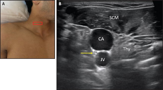
Figure 8-8. Vagus nerve. (A) Probe placement transversely over the sternocleidomastoid muscle at the level of the C6 anterior tubercle. (B) With the sternocleidomastoid muscle (SCM) as the most superficial structure and the thyroid gland (Thy) medially, the vagus nerve (yellow arrow) is visualized between the internal carotid artery (CA) and internal jugular vein (JV), inside the carotid sheath.
- Landmarks: With the sternocleidomastoid as the most superficial structure and the thyroid gland medially, the vagus nerve is visualized between the internal carotid artery and internal jugular vein, inside the carotid sheath. Lateral to the carotid artery are the longus capitis and longus colli muscles. The cervical ganglion can be seen between the longus capitis and longus colli muscles (Figure 8-9).4
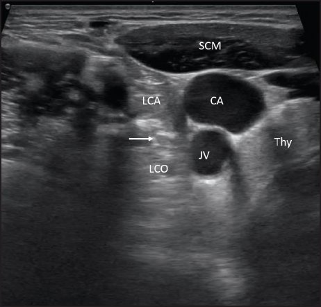
Figure 8-9. Cervical ganglion. Probe placement is the same as for the vagus nerve, with slightly lateral placement. Lateral to the carotid artery (CA) are the longus capitis (LCA) and longus colli (LCO) muscles. The cervical ganglion (white arrow) can be seen between them. (JV = jugular vein; SCM = sternocleidomastoid muscle; Thy = thyroid gland.)
- Relevant anatomy: The vagus nerve is the tenth cranial nerve with both motor and sensory fibers.5
- Points to remember: There are 3 cervical ganglia connected by intervening cords: superior, middle, and inferior. At the level of the C6 vertebra, the middle cervical ganglion can be seen; it receives ganglia contribution from C5 and C6.5
- Landmarks: With the sternocleidomastoid as the most superficial structure and the thyroid gland medially, the vagus nerve is visualized between the internal carotid artery and internal jugular vein, inside the carotid sheath. Lateral to the carotid artery are the longus capitis and longus colli muscles. The cervical ganglion can be seen between the longus capitis and longus colli muscles (Figure 8-9).4
- Probe/patient position: The patient’s head is rotated to the opposite side, and the probe is placed posteriorly on the sternocleidomastoid muscle in the slight sagittal oblique axis in the upper third of the muscle. The greater auricular nerve is visualized overlying the sternocleidomastoid muscle (Figure 8-10).4
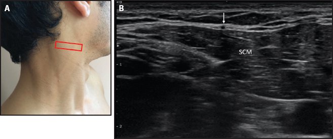
Figure 8-10. Greater auricular nerve. (A) Probe placement posteriorly on the sternocleidomastoid muscle in the slight sagittal oblique axis in the upper third of the muscle. (B) The greater auricular nerve (white arrow) is visualized overlying the sternocleidomastoid muscle (SCM).
- Landmark: Sternocleidomastoid muscle in the upper third of the muscle belly
- Relevant anatomy: The greater auricular nerve originates from the cervical plexus and is composed of the C2 and C3 spinal nerves. It emerges around the posterior border of the sternocleidomastoid muscle and ascends superiorly to give sensory innervation to the skin over the parotid gland, mastoid process, and outer ear.5
- Probe/patient position: The patient’s head is rotated to the opposite side, and the probe is placed on the sternocleidomastoid muscle in the sagittal oblique axis in the upper third of the muscle in a posterior-to-anterior orientation. The spinal accessory nerve is visualized within the belly of the sternocleidomastoid muscle (Figure 8-11).4
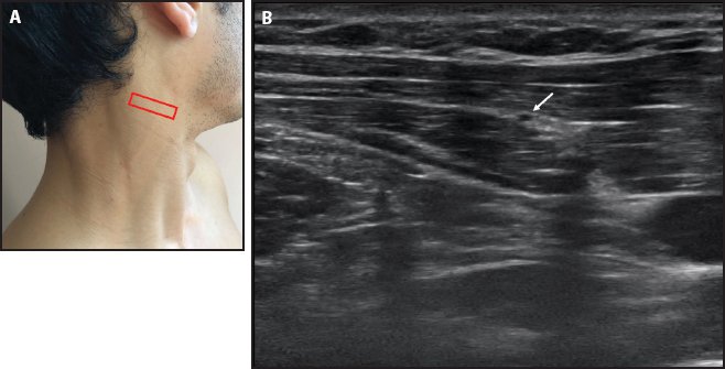
Figure 8-11. Spinal accessory nerve. (A) Probe placement on the sternocleidomastoid muscle in the sagittal oblique axis in the upper third of the muscle in a posterior-to-anterior orientation. (B) The spinal accessory nerve (white arrow) is visualized within the belly of the sternocleidomastoid muscle.
- Landmark: Sternocleidomastoid muscle in the upper third of the belly
- Relevant anatomy: The spinal accessory nerve is the 11th cranial nerve. It gives motor innervation to the sternocleidomastoid and trapezius muscles. It pierces the sternocleidomastoid muscle and courses obliquely across the posterior triangle of the neck. As it traverses the sternocleidomastoid muscle, it gives innervating branches to the muscle and is then joined by branches from the C2, C3, and C4 spinal nerve roots posteriorly to form a plexus that gives innervation to the trapezius muscle.5
- Probe/patient position: The patient’s head is rotated to the opposite side, and t he probe is placed in the sagittal oblique axis, posterior to the sternocleidomastoid muscle and over the middle scalene belly at the level of the C5 nerve root. The dorsal scapular nerve is visualized within the middle scalene muscle belly (Figure 8-12).4
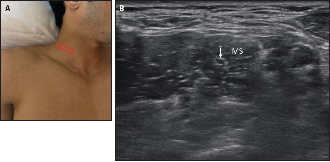
Figure 8-12. Dorsal scapular nerve. (A) Probe placement in the sagittal oblique axis, posterior to the sternocleidomastoid muscle and over the middle scalene belly at the level of the C5 nerve root. (B) The dorsal scapular nerve (white arrow) is visualized within the middle scalene (MS) muscle belly.
- Landmarks: Middle scalene muscle and C5 nerve root
- Relevant anatomy: The dorsal scapular nerve arises from the brachial plexus and has fiber contribution from the C5 nerve root. It gives motor innervation to the rhomboids and levator scapulae muscle. It pierces the middle scalene and then passes beneath the levator scapulae muscle as it courses posteriorly.3,5
- Probe/patient position: The patient’s head is rotated to the opposite side, and the probe is placed along the LX of the nerve just below the ear. The nerve is visualized in the LX going through the parotid gland (Figure 8-13).6
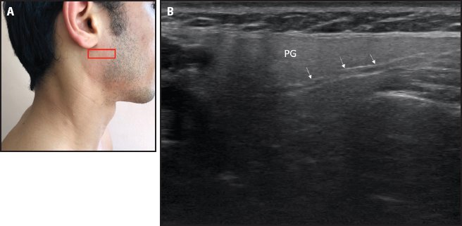
Figure 8-13. Facial nerve. (A) Probe placement. (B) The facial nerve (white arrows) is visualized in the LX going through the parotid gland (PG).
- Landmarks:
a. Internal: Parotid gland
b. External: Just below the ear
- Relevant anatomy: The facial nerve is the seventh cranial nerve. It exits the skull through the stylomastoid foramen and divides into 5 branches as it pierces through the parotid gland: temporal, zygomatic, buccal, mandibular, and cervical.
Greater and Lesser Occipital Nerves
- Probe/patient position:
a. Greater occipital nerve: The p robe is placed in an oblique axis between the spinous process of the C2 and mastoid process. The greater occipital nerve is visualized between the semispinalis capitis and obliquus capitis inferior muscles (Figure 8-14).4
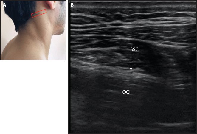
Figure 8-14. Greater occipital nerve. (A) Probe placement in the oblique axis between the spinous process of the C2 and mastoid process. (B) The greater occipital nerve (white arrow) is visualized between the semispinalis capitis (SSC) and obliquus capitis inferior (OCI) muscles.
b. Lesser occipital nerve: The patient’s head is rotated to the opposite side, and the probe is placed transversely on the posterior border of the sternocleidomastoid at the level of the upper third of the muscle, just below the hairline. The lesser occipital nerve is visualized between the sternocleidomastoid and levator scapulae muscles (Figure 8-15).
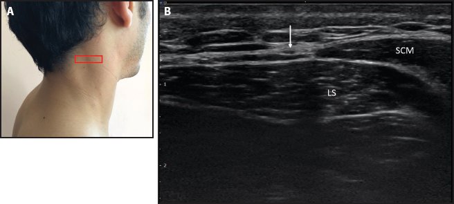
Figure 8-15. Lesser occipital nerve. (A) Probe placement transversely on the posterior border of the sternocleidomastoid muscle at the level of the upper third of the muscle, just below the hairline. (B) The lesser occipital nerve (white arrow) is visualized between the sternocleidomastoid (SCM) and levator scapulae (LS) muscles.
- Landmarks:
a. Greater occipital nerve: The spinous process of the C2 and mastoid process are external landmarks. The semispinalis capitis and obliquus capitis inferior muscles are internal landmarks.
b. Lesser occipital nerve: The posterior border of the sternocleidomastoid and levator scapulae muscles. The levator scapulae muscle is lined by the hyperechoic deep cervical fascia.
- Relevant anatomy: The greater occipital nerve arises from the medial branch of the dorsal ramus of the C2. The lesser occipital nerve arises from the ventral ramus of the C2.
- Landmarks:
- Probe/patient position: The patient’s head is rotated to the opposite side, and the p robe is placed on the sternocleidomastoid muscle at the level of the mid-belly. The phrenic nerve is visualized superficial to the anterior scalene muscle and deep to the thyrocervical trunk of the subclavian artery (Figure 8-16).4
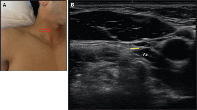
Figure 8-16. Phrenic nerve. (A) Probe placement on the sternocleidomastoid muscle at the mid-belly level. (B) The phrenic nerve (yellow arrow) is visualized superficial to the anterior scalene muscle (AS).
- Landmarks:
a. External: Posterior border of the sternocleidomastoid muscle at the mid-belly level
b. Internal: Anterior scalene muscle and thyrocervical trunk of the subclavian artery and sternocleidomastoid muscle
- Relevant anatomy: The phrenic nerve originates from the anterior division of C3, C4, and C5. It receives contribution from both the cervical plexus and brachial plexus.
- Probe/patient position: The patient’s head is rotated to the opposite side, and the p robe is placed above and along the clavicle. The subclavian nerve is visualized just above the subclavian artery. Immediately next to it, the brachial plexus cluster can be visualized (Figure 8-17).4
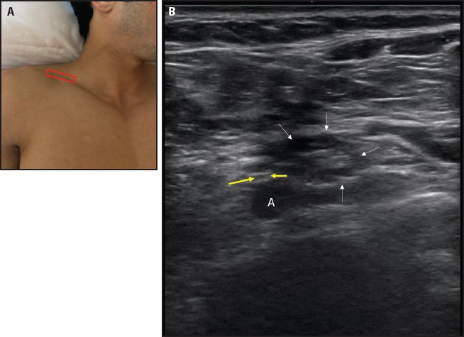
Figure 8-17. Subclavian nerve. (A) Probe placement above and along the clavicle. (B) The subclavian nerve (between the yellow arrows) is visualized just above the subclavian artery (A) and immediately next to the brachial plexus cluster (white arrows).
- Landmarks:
a. External: Clavicle
b. Internal: Subclavian artery and brachial plexus
- Relevant anatomy: The subclavian nerve originates from the junction point of the C5 and C6 nerve roots. It is connected by a filament with the phrenic nerve.5
- Probe/patient position: The probe is placed in the SX over the middle scalene muscle at the level of the C5 and C6 nerve root. The long thoracic nerve is visualized within the middle scalene muscle, posterior to the C5 and C6 nerve roots (Figure 8-18).4
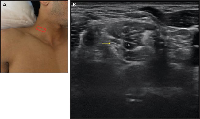
Figure 8-18. Long thoracic nerve. (A) Probe placement in the SX over the middle scalene muscle at the level of the C5 and C6 nerve roots. (B) The long thoracic nerve (yellow arrow) is visualized within the middle scalene muscle, posterior to the C5 and C6 nerve roots.
- Landmarks: Middle scalene muscle and C5 and C6 nerve roots
- Relevant anatomy: The long thoracic nerve arises from the C5, C6, and C7 nerve roots and supplies the serratus anterior muscle.5
- Probe/patient position:
a. Suprascapular notch: The probe is positioned over the suprascapular notch and aligned along the direction of the axis of the coracoid process. The suprascapular nerve is visualized in the suprascapular notch with the superior transverse ligament overlying it and the suprascapular artery superficial to the ligament (Figure 8-19).
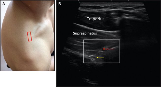
Figure 8-19. Suprascapular nerve from the suprascapular notch. (A) Probe placement over the suprascapular notch, with the probe aligned along the direction of the axis of the coracoid process. (B) Suprascapular nerve (yellow arrow) and artery (red arrow).
b. Spinoglenoid notch: The probe is placed over the posterior glenohumeral joint and then moved medially and rotated slightly in the SX to the spinoglenoid notch area. The suprascapular nerve is visualized with the suprascapular artery in the notch (Figure 8-20).7
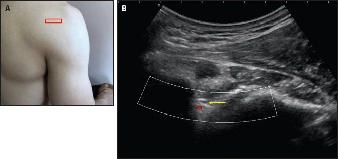
Figure 8-20. Suprascapular nerve from the spinoglenoid notch. (A) Probe placement over the posterior glenohumeral joint, then moved medial and rotated slightly to the SX view of the spinoglenoid notch area. (B) Suprascapular nerve (yellow arrow) and artery (red area).
- Landmarks: Suprascapular notch and coracoid process (for probe orientation); spinoglenoid notch and posterior glenohumeral joint
- Relevant anatomy: The supraspcapular nerve arises from the superior trunk of the brachial plexus and has fibers derived from the C5 and C6 nerve roots. It gives motor supply to the supraspinatus and infraspinatus muscles and sensory innervation to the acromioclavicular joint, subacromial bursa, and glenohumeral joint.
- Points to remember: It is important to turn on color Doppler mode to identify the artery, which helps in visualization of suprascapular nerve. The artery is superficial to the superior transverse ligament in the suprascapular notch. The artery accompanies the suprascapular nerve in the spinoglenoid notch.
- Landmarks: Suprascapular notch and coracoid process (for probe orientation); spinoglenoid notch and posterior glenohumeral joint
- Probe/patient position: The posterior cord from the subpectoralis minor space is followed laterally to visualize the axillary nerve between the subscapularis and coracobrachialis/short head of the biceps muscle (Figure 8-21). The probe is placed in the LX on the posterior aspect of the shoulder at the level of the teres minor muscle. The axillary nerve (posterior branch) is seen between the teres minor and the lateral head of the triceps, overlying the humeral bony interface and accompanied by the posterior circumflex humeral artery (Figure 8-22).1
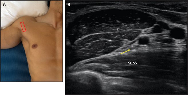
Figure 8-21. Axillary nerve anteriorly. (A) Probe placement. The posterior cord of the subpectoralis minor space is followed laterally to visualize the axillary nerve between the subscapularis and coracobrachialis/short head of the biceps muscle. (B) The axillary nerve (yellow arrow) is seen between the subscapularis (SubS) and coracobrachialis/short head of the biceps muscle (small white B).
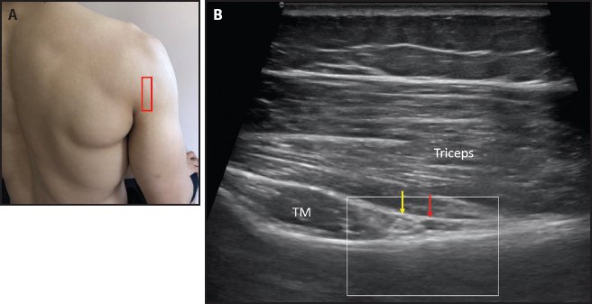
Figure 8-22. Axillary nerve posteriorly. (A) Probe placement in the LX on the posterior aspect of the shoulder at the level of the teres minor muscle. (B) The axillary nerve posterior branch (yellow arrow) is seen between the teres minor (TM) and lateral head of the triceps, overlying the humeral bony interface and accompanied by the posterior circumflex humeral artery (red arrow).
- Landmarks: The teres minor muscle, lateral head of the triceps, and posterior circumflex artery are internal landmarks.
- Relevant anatomy: The axillary nerve arises from the posterior cord of the brachial plexus and derives fibers from the C5 and C6 roots.
Ulnar, Median, Radial, and Musculocutaneous Nerves
- Probe/patient position: The probe is placed in the oblique LX along the anterior axillary fold, and the axillary artery is identified. With respect to the axillary artery, the median nerve is lateral, the ulnar nerve is medial, and the radial nerve is posterior (Figure 8-23). Laterally, the musculocutaneous nerve is seen between the biceps brachii and coracobrachialis muscles (Figure 8-24).3
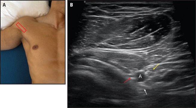
Figure 8-23. Median, ulnar, and radial nerves at the anterior axillary fold. (A) Probe placement. (B) Median (yellow arrow), ulnar (white arrow), and radial (red arrow) nerves around the axillary artery (white A).

Stay updated, free articles. Join our Telegram channel

Full access? Get Clinical Tree


