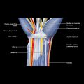
Peritendinous Mass
ESSENTIAL INFORMATION
Key Differential Diagnosis Issues
 Only some tendons, notably those around wrist and ankle, have tendon sheaths
 Some tendon areas, such as patellar and Achilles tendons, have incomplete tendon sheaths known as paratenon
 Some tendon areas, such as extensor tendons at fingers and flexor capri ulnaris tendon, do not have tendon sheath
 Tenosynovitis and giant cell tumor of tendon sheath can only occur in tendon with tendon sheath or paratenon
Helpful Clues for Common Diagnoses
 Filled with gelatinous-type material of variable consistency
 Anechoic, cystic-like structure closely related to joint
 Usually originates from joint and extends to peritendinous location
 If originating from tendon sheath, has no extension to adjacent joint
 ± comet-tail artifacts within cyst due to colloid aggregates
 ± leakage of or hemorrhage within cyst
 Composed of multinucleated cells and fibroblast-like cells that may have hemosiderin deposits
 Occurs mainly in hand, wrist, and foot
 All giant cell tumors contact or partially encase tendon
 Hypoechoic tumor, which may be irregular, fusiform, or rounded in outline
 Usually moderately vascular, though, infrequently no demonstrable flow on color Doppler imaging
 May be multifocal
 High recurrence rate
 Can mimic ganglion cyst
 Inflammation or infection of tendon sheath with secondary inflammation of tendon
 Acute exudative tenosynovitis
 Acute nonexudative tenosynovitis
![]()
Stay updated, free articles. Join our Telegram channel

Full access? Get Clinical Tree


