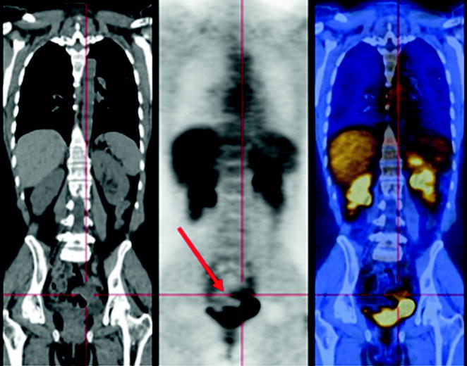
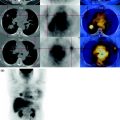
Fig. 28.1
The small pelvis in correspondence of the bladder trigone in the left paramedian region (See description in the text (Report))
Absence of pathological glucose consumption in the remaining parts of the body examined.
28.4 Conclusions
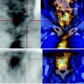
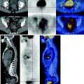
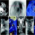
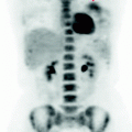
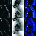
The PET scan shows slight increase in glucose metabolism reported for peritoneal carcinomatosis due to pelvic lesion and the lesion on the splenic surface (See Figs. 28.2, 28.3).
