PET and PET-CT of the Liver and Pancreas
Thomas F. Hany
Klaus Strobel
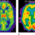
The imaging and differentiation of primary solid liver tumors is a domain of morphological imaging. In fluorodeoxyglucose (FDG) positron emission tomography (PET) imaging, well-differentiated hepatocellular carcinomas as well as all benign solid liver tumors do not show increased FDG uptake. Therefore, PET may only be useful for differentiating benign solid liver tumors from liver metastases in cancer patients if radiological imaging procedures fail. On the other hand, in patients with known moderately to poorly differentiated hepatocellular carcinoma or cholangiocarcinoma, PET imaging has been shown to be useful, especially in the detection of distant metastases and follow-up after treatment (see Fig. 42.1). Other applications of PET in primary infectious liver disease include therapy monitoring of echinococcus infection.
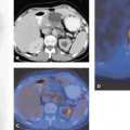
In pancreatic disease, computed tomography (CT) and magnetic resonance imaging (MRI) are the cornerstones of diagnosis and staging, because both modalities can exactly delineate the vascular and adjacent structures. However, the differentiation of pancreatic masses into chronic pancreatitis or pancreatic carcinoma remains difficult. Initial results from the use of FDG-PET-CT in the presurgical evaluation of pancreatic masses are promising, and the combination of FDG-PET-CT, fine needle aspiration, and tumor markers seem to be cost-efficient in the management pancreatic cancer. In the future, using PET-CT, with the inclusion of dynamic kinetics, delayed FDG imaging, and dedicated contrast-enhanced CT in the same imaging session, could possibly evolve into the modality of choice.
Introduction
Efforts have been made to differentiate benign from malignant liver lesions by morphological and functional imaging modalities. Since most of the tumors found are solid and therefore not easily characterized, additional morphological characteristics have been defined to differentiate benign from malignant lesions using form of the lesion, tissue densities, flow patterns, contrast enhancement over time, and signal intensities. Nevertheless, in many cases histological workup is still needed for definite diagnosis, especially in patients with known cancer.
In the recent years, use of fluorodeoxyglucose (FDG) positron emission tomography (PET) as an adjunct to morphological imaging modalities has increased. The ability to measure the glucose consumption of any kind of tissue by PET and therefore possibly characterize these lesions has attracted the attention of many clinicians. In general, malignant lesions show a higher FDG accumulation due to FDG trapping in tumor cells, which can be explained by decreased intracellular glucose-6-phosphatase (G-6-P) and increased hexokinase (HK) concentrations compared with normal hepatocytes (1). Additionally, this misbalance could also be directly correlated to the grade of tumor aggressiveness (1). Unfortunately, well-differentiated tumor cells may show normal or only slightly increased FDG uptake in relation to surrounding hepatocytes. Additionally, inflammation shows also increased FDG uptake, which is of major concern in the evaluation of pancreatic masses, when malignant disease has to be excluded (2). To overcome this problem, new data-acquisition strategies like dynamic kinetics and delayed imaging in FDG-PET imaging have been developed (3).
Liver
Infectious Diseases of the Liver
Liver abscess and other infectious diseases, such as echinococcus disease, can be detected by FDG-PET (4,5,6). Unfortunately, those lesions may be indistinguishable from metastatic disease or primary liver tumors. Further, increased FDG uptake in the intrahepatic bile ducts after interventional therapy interferes with diagnosis. FDG-PET is not suited to be a first-line imaging modality in the detection of infectious disease of the liver but may be useful for follow-up examinations in selected diseases, such as in the measurement of disease activity in echinococcus disease (5).
Benign Primary Liver Tumors
Hemangiomas are benign liver lesions that occur in 4% and 7% of the population and may enlarge, especially during increased estrogen levels (pregnancy, iatrogenic). The typical hemangioma is small, asymptomatic, and discovered incidentally. Giant hemangiomas (diameter greater than 5 cm) may become symptomatic due to hemorrhage and thrombosis. Typical radiomorphologic features include hyperechoic lesions in ultrasound; hypodense, well-circumscribed lesions in non-contrast-enhanced CT; and nodular peripheral enhancement in contrast-enhanced CT scans. The imaging modality of choice is magnetic resonance imaging (MRI) by using heavily T2-weighted data acquisition, which demonstrate a typical hyperintense lesion (light bulb sign). Technetium 99m (99mTc)-labeled red blood cells have been used and show a typical increased accumulation during the blood pool phase in delayed imaging, but this technique has been superseded by MRI. FDG-PET imaging shows normal or decreased FDG uptake after 40 to 90 minutes. However, differentiation between tumor necrosis or inflammation by PET imaging is difficult, and therefore PET is not the imaging modality of choice.
Focal nodular hyperplasia (FNH) is rare benign hepatic neoplasm that is mostly seen in young female patients and has no risk of malignant transformation. Tumor tissue includes hepatocytes, bile duct elements, Kupffer cells, and collagenous tissue, which imposes as hypervascular structure. A central fibrous scar is common. [99mTc]sulfur colloid uptake is increased in 70% of the cases. FDG-PET imaging of such lesions shows no increased FDG uptake (7). Therefore, PET imaging is nonspecific and may be only useful for differentiating FNH from liver metastases in cancer patients if radiological imaging procedures fail.
Liver adenomas are less common than FNH, composed only of hepatocytes, and associated with oral contraceptives and glycogen storage disease. Radiological features include hypodense lesions in CT and variable echo-structure in ultrasound. Adenomas show no more uptake of [99mTc]sulfur colloids than does FNH. FDG-PET imaging has been described as showing no significant difference in uptake between adenomas and normal hepatic tissue (8).
In benign cystic lesions, FDG-PET shows decreased FDG uptake. However, only larger lesions (greater than 1 to 2 cm) may be seen as signal void in PET imaging. Therefore, FDG-PET imaging in benign solid hepatic lesions may only be useful for differentiating unclear radiological findings from liver metastases in cancer patients.
Malignant Primary Liver Tumors
Malignant primary liver tumors include hepatocellular carcinoma, fibrolamellar carcinoma, cholangiocarcinoma of the intrahepatic bile ducts, and mixed hepatocellular carcinoma and cholangiocarcinoma. Hepatic carcinomas are frequently multifocal and have been shown to involve multiple sites in both liver lobes at the time of exploration. Preoperative assessment should also include a search for extrahepatic metastases, since this condition will preclude a curative approach.
Hepatocellular carcinoma (HCC) is the most common primary visceral malignancy worldwide. The natural history varies widely among global regions, with the highest incidences found in southern Africa and Asia (especially Japan) (5% to 20%). Etiological factors and cofactors include chemical carcinogens (aflatoxin) and cirrhosis, which is irrespective of the etiology. However, cirrhosis induced by hepatitis B and C is a common cause. Carcinogen-related HCC grows fast in normal liver tissue and slower in cirrhotic livers. Solitary, multiple, and diffuse forms are described. Measurement of α-fetoprotein (AFP) may be helpful in diagnosis and as a prognostic factor. Several histological variants are described, and fibrolamellar carcinomas have the best prognosis if diagnosed early. However, the median survival of untreated patients is poor and ranges from 1 to 4 months after diagnosis. Interestingly, extrahepatic metastasis of HCC occurs relatively late in the clinical course and therefore does not significantly shorten the survival time. Whenever the stage of disease allows, surgical treatment should be pursued (9,10).
The diagnostic workup of hepatic lesions includes ultrasound, dynamic contrast-enhanced CT, and MRI. Ultrasound has a rather low sensitivity but a high specificity in detecting HCC. It is used beneficially in screening programs for HCC (11). In CT imaging, lesions appear as an imposing hypodense mass with early arterial enhancement. An advantage of this imaging modality is that it allows the simultaneous evaluation of vascular structures, such as the hepatic and portal veins, and their relationship to the tumor. Further diagnostic workup may include CT arterial portography (CTAP), which is an invasive procedure, since an intra-arterial catheter needs to be placed into the visceral arteries. However, the recently introduced
multi-row helical CT seems to produce equivalent results without invasive procedures such as needed for CTAP (sensitivity, 94%; positive predictive value, 96%) (12).
multi-row helical CT seems to produce equivalent results without invasive procedures such as needed for CTAP (sensitivity, 94%; positive predictive value, 96%) (12).
Stay updated, free articles. Join our Telegram channel

Full access? Get Clinical Tree


