| SKULL BASE REGION | Retromeatal petrosal surface |
| HISTOPATHOLOGY | Meningioma, transitional |
| PRIOR SURGICAL RESECTION | Yes |
| PERTINENT LABORATORY FINDINGS | N/A |
Case description
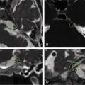
The patient is a 58-year-old female who, 20 years prior to presentation, underwent surgical resection of a transitional meningioma of the right petrous bone extending into the sigmoid sinus and tentorium. An asymptomatic, small, left lesser sphenoid wing meningioma was also detected at that time. The patient was lost to follow-up until December 2018. She then underwent brain magnetic resonance imaging (MRI), which showed growth of both the residual tumor invading the sigmoid sinus and the middle cranial fossa meningioma ( Figure 7.32.1 ). Her only symptom was tinnitus in the right ear with normal hearing. She underwent multisession CyberKnife stereotactic radiosurgery (SRS) for both meningiomas over 2 consecutive weeks ( Figure 7.32.2 ).
| Radiosurgery Machine | CyberKnife |
| Radiosurgery Dose (Gy) | 25, at the 80% isodose line |
| Number of Fractions | 5 |

Preoperative postcontrast T1-weighted image showing the remnant tumor invading the right sigmoid sinus and crossing through the tentorium as well as the left lesser sphenoid wing tumor, abutting the cavernous sinus and extending to the middle cranial fossa.

Imaging of the treatment plan.
| Critical Structure | Dose Tolerance |
|---|---|
| Sigmoid sinus | 25–27 Gy in 5 fractions (internal guideline) |
| VIII cranial nerve | 25 Gy in 5 fractions (internal guideline) |
Related posts:
 Esthesioneuroblastoma – delayed postoperative radiosurgery for recurrence at long-term
 Null cell – delayed postoperative radiosurgery for growing perioptic residual
 Chondrosarcoma – definitive radiosurgery after subtotal resections
 Trigeminal neuralgia due to microvascular conflict – upfront radiosurgery
 Capillary hemangioma – postoperative radiosurgery for residual tumor
 Superior sagittal sinus meningioma – delayed postoperative, multisession radiosurgery for growing residual
Stay updated, free articles. Join our Telegram channel

Full access? Get Clinical Tree


