A. Shorter exposure time
B. Increased spatial resolution
C. Decreased glandular dose
D. Increased contrast
E. Decrease density
ANSWERS AND EXPLANATIONS
1 Answer A. Grids are used routinely in mammography to increase image contrast. Most mammography systems have a moving grid with a ratio of 4:1 to 5:1 focused to the source to image distance. Grids do not compromise spatial resolution, but they do increase patient dose. However, the dose is still acceptably low, and the improvement in contrast is significant.
Reference: Bushong SC. Radiologic Science for Technologists: Physics and Protection. 10th ed. St. Louis, MO: Elsevier Mosby; 2013:381.
2 Answer C. Breast compression results in decreased tissue thickness. The scatter to primary ratio for a compressed breast is 0.4 to 0.5, whereas the scatter to primary ratio for a noncompressed breast is 0.8 to 1.0. Reducing tissue thickness allows for use of lower mAs, which results in decreased radiation dose. Compression results in reduced exposure dynamic range because tissue is spread out creating a more uniform thickness. Magnification can be produced with an air gap.
Reference: Bushberg JT, Seibert JA, Leidholt EM, et al. The Essential Physics of Medical Imaging. 2nd ed. Philadelphia, PA: Lippincott Williams & Wilkins; 2001:207.
3 Answer C. A very good, short white paper on mammography reading room design is Albert Xthona, “Designing the Perfect Reading Room for Digital Mammography,” Barco White Paper, 2003 and is available online at this link: http://www.barco.com/barcoview/downloads/The_perfect_mammography_reading_room_2011_-_White_paper.pdf. This is the primary resource for this question. The paper shows a long narrow room with all workstations along one wall, emphasizing the need to reduce light falling from one monitor to another. Thus, all the workstations are in a straight line.
A. Though workstations are sometimes placed angled slightly toward each other; this greatly increases the background lighting at each monitor and substantially decreases the image contrast.
B. Walls should be dark colored to reduce the ambient light. Light for walking safely should be very near the ground, with low-level lamps placed below the desks and pointing toward the floor.
D. Mammography view boxes are sometimes used as a source of light for writing or moving about the room. This is not a good idea, and the reference shows how to better accommodate these needs.
4 Answer D. This covers the range of target/filter combinations used in digital mammography, and the tungsten target + silver filter (k edge energy of 25 keV) will have the highest energy and greatest penetrating power.
Reference: Huda W. Review of Radiographic Physics. 3rd ed. Baltimore, MD: Lippincott Williams & Wilkins; 2010:51–52.
5 Answer E. Moving grids (Bushong, p. 200ff) are used in contact mammography (but not in magnification mammography) solely for the reduction of scatter to the image receptor. Note that the Mammography Quality Safety Act (MQSA) requires [900.12(b)(4)] that
1 (ii) Systems using screen-film image receptors shall be equipped with moving grids matched to all image receptor sizes provided.
2 (iii) Systems used for magnification procedures shall be capable of operation with the grid removed from between the source and image receptor.
Grids are generally used for the same reasons for machines with digital receptors as well. Breast compression has many benefits including the reduction of scatter (Bushong, p. 380).
Answer choice A is wrong because “compression results in thinner tissue and therefore less scatter radiation (Bushong, p. 380). ” Scatter is higher at higher kV (Bushong, pp. 187, 188) and so answer choice B is wrong. Answer choice C is a little tricky. Breast compression reduces the scatter and thus the benefit of scatter reduction using a grid, but, in that, “the use of grids during (contact) mammography is routine (Bushong, p. 381),” the grid still provides enough benefit to warrant its use and so answer choice C is wrong. This subject is explored further in Gennaro et al. for magnification mammography; however, the air gap sufficiently reduces the effects of scatter so that the grid is not used (Bushberg et al., p. 210) and so answer choice D is incorrect.
References: Bushong SC. Radiologic Science for Technologists. 10th ed. St. Louis, MO: Elsevier; 2013.
Bushberg JT, Seibert JA, Leidholdt EM, et al. The Essential Physics of Medical Imaging. 2nd ed. Philadelphia, PA: Lippincott Williams & Wilkins; 2001.
Gennaro G, Katz L, Souchay H, et al. Grid removal and impact on population dose in full-field digital mammography. Med Phys 2007;34(2):547–555.
MQSA: http://www.fda.gov/Radiation-EmittingProducts/MammographyQualityStandardsActand Program/Regulations/ucm110906.htm
6 Answer C. Many of the points in the discussion below are taken from an excellent tutorial on mammographic displays by Ehsan Samei.
Many of the quantitative terms (and there are many) used in optics have evolved over centuries of use and have a quaint, even charming, but often confusing feel to them. Luminance and illuminance are two of these terms. While both now have official SI definitions, they still bear a relationship to their everyday English meanings. Luminance is the perceived brightness of a display and can apply to both view boxes and the monitors used for digital display. (Illuminance describes the outside light falling on the display and while illuminance is good when reading a book, it degrades the images from monitors used for mammographic displays by reducing the perceived contrast.) Now on to the question.
The answer choice A is wrong because there is a limit to the brightness range comfortable (and at extremes, safe) for the human vision system. (That is why we have sunglasses, for example.) Answer choice B is somewhat better but again, as we go too high in the contrast ratio displayed, we again reach the limits of the adaption capabilities of the human visual system. We also run into problems due to the contrast reduction processes of veiling glare and reflection. The answer choice D is incorrect because even the best mammographic film cannot compete with the dynamic range in contrast of modern digital displays. (Film still beats digital displays in resolution, however. That is it can see tiny objects of high contrast better than digital displays.)
Answer choice C is correct. Modern digital displays can easily, as previously stated, exceed the contrast display capabilities of film so a good minimum contrast ratio is that of film on a standard view box. However, it should not be too high for the reasons given above in discussing answer choice B.
Reference: Samei E. Technological and psychophysical considerations for digital mammographic displays. Radiographics 2005;25:491–501.
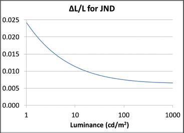
7 Answer A. Imagine looking at a display with a uniform background of brightness L. The display is divided into two sections but initially both are matched in brightness so that they appear as one. Now imagine that one side (e.g., the left side) is very slowly made brighter while you are viewing the display. At some point you perceive that there are two sections with the left half just barely brighter than the right half. This difference in brightness ΔL is just noticeable and so it is called the just noticeable difference (JND). Dividing the JND by the luminance itself is called the contrast threshold. In other words, the contrast threshold is the fractional change in luminance ΔL/L required to be just noticeable. If the contrast threshold was a constant, independent of the luminance value, then Weber law would strictly hold for visual contrast perception. It is not true but does provide us with a good starting point. (Weber law is a rough approximation which can be applied to a number of sensory modalities, e.g., sound loudness perception.)
The plot of the gray-scale standard display function taken from the DICOM 14 document (http://medical.nema.org/dicom/2004/04_14pu.pdf) shows that for dimly luminated displays, a larger contrast is required to be perceptible than for brighter displays and so Weber law does not hold. For brighter screens (above 100 cd/m2), the curve flattens out to the right edge of the displayed range and beyond. (A bright mammography viewing box is 3,000 cd/m2) This is the situation described in the correct answer choice A.
References: Samei E. Technological and psychophysical considerations for digital mammographic displays. Radiographics 2005;25:491–501.
8 Answer D. Note that mean glandular dose (MGD) is the average dose throughout the breast. The dose at the beam entrance surface of the breast would be the highest. As the beam passes through the breast, it is attenuated and so the dose decreases as it passes through the breast. Thus, the average dose is lower than the dose near the beam entrance.
The problem states that the entrance exposure has increased by 60% with a 16% increase in kVp. In addition, the penetrability (range of x-rays in tissue) goes up with kVp (Bushong, p. 140). (This is equivalent to saying that the x-ray attenuation is less at higher kVp.) What effect does this have? It means that as the radiation passes through the tissue, it does not get reduced as much as it would at lower kVp. Thus, the mean (average) dose to the tissue is higher than it would have been had the penetrability remained constant. Thus, the dose increases by more than 60%.
It is important to note that according to the problem, the mAs has been held constant. Normally, if one chooses to increase the kVp, the system would automatically reduce the mAs.
Reference: Bushong SC. Radiologic Science for Technologists. 10th ed. St. Louis, MO: Elsevier; 2013.
9 Answer B. CsI (indirect) is used by GE; Se (direct) is used by Hologics. BaFBr is used by FUJI (photostimulable phosphor). NaI is used in gamma cameras, not mammography.
Reference: Huda W. Review of Radiographic Physics. 3rd ed. Baltimore, MD: Lippincott Williams & Wilkins; 2010:53.
10 Answer B. In conventional radiography (80 kV), about 2.5 to 3 cm of soft tissue attenuates half the x-ray beam; in mammography, about 1 cm of soft tissue will reduce the primary x-ray beam intensity to one half of its initial value.
Reference: Huda W. Review of Radiographic Physics. 3rd ed. Baltimore, MD: Lippincott Williams & Wilkins; 2010:54.
11 Answer C. Currently, in digital mammography, the typical x-ray tube voltage is 30 to 32 kV, with the higher value used in thicker breasts. Twenty-five kV would be far too low, and 40 kV would be far too high.
Reference: Huda W. Review of Radiographic Physics. 3rd ed. Baltimore, MD: Lippincott Williams & Wilkins; 2010:54.
12 Answer C. Tube currents are 100 mA for the large (0.3-mm focal spot) and only 25 mA for the small focal spot (0.1 mm); as a result, to get a given mAs in magnification mammography, the exposure time must be increased (threefold) to get the correct exposure at the image receptor.
Reference: Huda W. Review of Radiographic Physics. 3rd ed. Baltimore, MD: Lippincott Williams & Wilkins; 2010:54.
13 Answer C. mAs is an abbreviation for milliampere second, the units of measure of x-ray tube current and exposure time (Bushong, p. 593). Thus, at fixed mAs, if the exposure time goes up, the tube current must go down. At constant kVp, the dose to the breast is proportional to the mAs, which has not changed. (It has just been delivered to the breast over a longer period of time but at a lower rate.) Thus, answer choice A is incorrect. There would also be no difference in the total amount of radiation impinging on the detector and so the viewing requirements should not change. Thus, answer choice B is also incorrect. Answer choice C is the correct answer since motion blur increases with exposure time (Bushong, pp. 181, 182). A fixed mAs means that for a longer exposure time, the tube current is reduced. Thus, answer choices D and E are both wrong. If anything, since the tube current and radiation rate have both been reduced, the exposure is gentler on both the tube and the detector.
Reference: Bushong SC. Radiologic Science for Technologists. 10th ed. St. Louis, MO: Elsevier; 2013.
14 Answer C. Calcium (Ca) has an atomic number of 20, whereas soft tissue has an effective atomic number of about 7.4 to 7.6. Though the attenuation coefficient drops with increasing keV for both calcifications (answer choice C) and normal breast tissue (answer choice D), the photoelectric effect for calcium is affected far more, and so the contrast between microcalcifications and normal breast tissue drops substantially as the photon energy increases (see Wolbarst, Fig. 33.3). Thus, the correct answer is C. Answer choice A is incorrect even though the large region of coverage for the chest radiograph might imply a decrease in resolution. Even with high resolution, we still would probably not see calcium in a chest x-ray because of the high kV. Answer choice B might be true except that the spine would generally not obfuscate the breasts in a chest radiograph and so this answer is easily eliminated. The range of exposure times can substantially overlap for mammograms and chest x-rays. Quite often, the exposure time will be shorter for a chest x-ray than for a mammogram and thus choice E is incorrect.
Reference: Wolbarst A. Physics of Radiology. 2nd ed. Madison, WI: Medical Physics Publishing Corp.; 2000:355–356.
15 Answer D. Because of the air gap in magnification mammography, much of the scatter from the breast does not reach the detector. Thus, there is little scatter reduction that can be accomplished by using a grid. Yet the grid, if present, would still attenuate a significant fraction of the primary beam, requiring an increase in breast dose for the same exposure to the detector. Thus, in magnification mammography, the grid is left out since it does little good and requires more dose to the breast. (None of this is unique to mammography.) Grids are used in contact (nonmagnification) mammography and so answer choice A is wrong. If the grids were present, it would be moving just as it does in contact mammography and so would be just as invisible as it would be in contact mammography. Thus, answer choice B is wrong. The contrast really should not be affected by magnification and so answer choice C is wrong. Answer choice E is more or less hogwash.
Reference: Bushberg JT, Seibert JA, Leidholdt EM, et al. The Essential Physics of Medical Imaging. 2nd ed. Philadelphia, PA: Lippincott Williams & Wilkins; 2001:207–212.
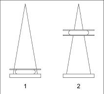
16 Answer B. Consider the geometry used for magnification mammography compared to imaging with the breast in contact with the detector. Start with the contact mammography depicted in Figure 5.1. The breast is compressed and in close contact with the detector. X-rays must penetrate through the compressed breast. As they pass through the breast, a fraction is absorbed (resulting in radiation dose to the breast.) After a sufficient amount of time, the detector receives enough radiation for an image and the automatic exposure control (AEC) turns the radiation off.
Stay updated, free articles. Join our Telegram channel

Full access? Get Clinical Tree


