Figure 1-72. Multipennate subscapularis muscle. [A] Short-axis 12-5 MHz US image shows normal multipennate subscapularis muscle (arrowheads). [B] Corresponding sagittal T1-weighted MRI confirms normal anatomy (arrowheads).

Figure 1-73. Normal multilaminar arrangement of the supraspinatus tendon. [A] Positioning of the probe. [B] Corresponding 12-5 MHz US image depicts a prominent layer of the supraspinatus tendon showing normal fibrillar pattern (arrowhead). This is a normal finding and should not be confused with calcification or fibrosis. Hum= humerus. Delt= deltoid.

Figure 1-74. Rotator cable. [A] Long-axis 12-5 MHz US image shows the normal rotator cable (arrowheads). [B] Corresponding coronal oblique T1-weighted MRI confirms the normal finding (arrowhead). Hum= humerus. Delt= deltoid.

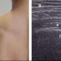
Figure 1-75. Normal musculotendinous junction. [A] Long-axis 12-5 MHz US image shows the normal transition between hyperechoic tendon (arrowhead) and hypoechoic muscle (asterisk) at the supraspinatus musculotendinous junction. [B] Corresponding coronal oblique STIR MRI confirms normal tendon (arrowheads). Hum= humerus. Delt= deltoid.
Related posts:

Stay updated, free articles. Join our Telegram channel

Full access? Get Clinical Tree


