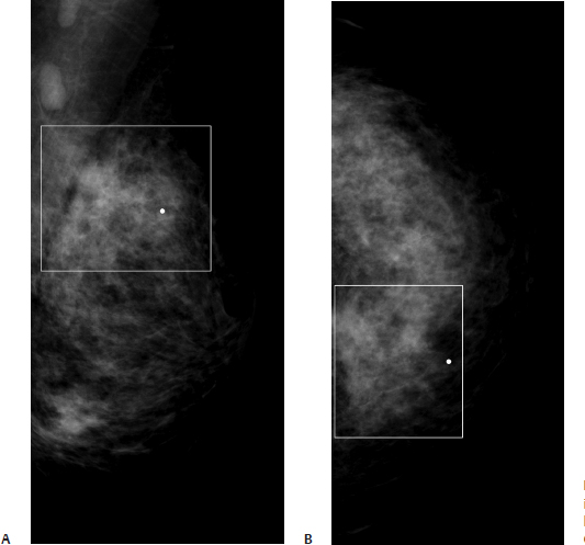
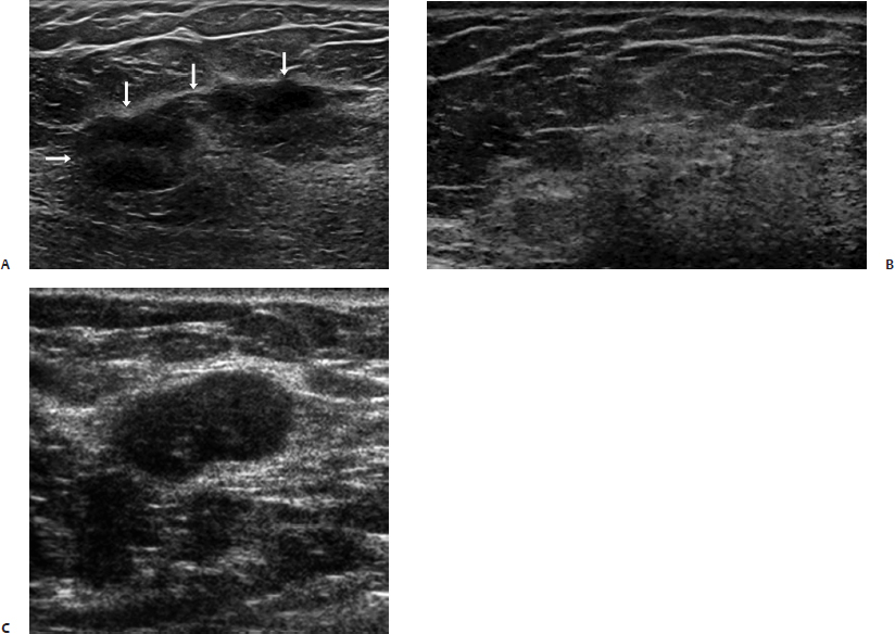
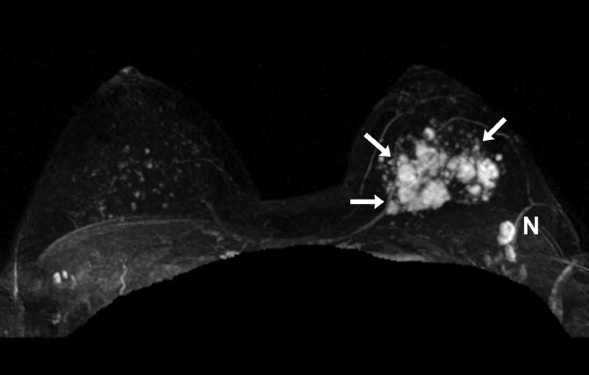
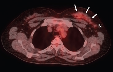
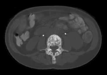
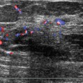
40 Pitfalls of PET A 54-year-old woman presents with left breast pain. • Left upper inner quadrant is diffusely tender. • Asymmetry in the upper inner quadrant • Abnormally dense lymph nodes Fig. 40.1 There is asymmetry in the left upper inner quadrant (square) associated with abnormal lymph nodes. (A) Left ML mammogram. (B) Left CC mammogram. Frequency (Fig. 40.2) • 15 MHz Fig. 40.2 The patient’s tenderness and mammographic asymmetry sono-graphically correspond to a left irregular hypoechoic mass (arrows) (A). Because the mass is large and ill-defined, the imager may easily miss the mass unless the hyperechoic normal adjacent glandular tissue (B) is compared with the hypoechoic mass. Sonography also demonstrates abnormal lymph nodes that lack a central hilum (C). (A) Left breast sonogram upper outer quadrant. (B) Left breast sonogram normal glandular tissue adjacent to mass. (C) Left axillary sonogram. Fig. 40.3 Contrast-enhanced bilateral breast maximum projection intensity (MIP) MRI demonstrates an enhancing, irregular, highly suspicious mass (arrows) and a lymph node (N) suspicious for metastatic disease. Fig. 40.4 Axial positron emission tomography–computed tomography (PET-CT). The area of MRI enhancement demonstrates only mild diffuse uptake (arrows) with maximum standardized uptake value (SUV) of 1.8. The enhancing lymph node (N) at the tail of the mass has a maximum SUV of 1.6. • Primarily invasive lobular carcinoma with a smaller proportion of invasive ductal carcinoma and ductal carcinoma in situ (DCIS). Axillary nodes were positive for metastatic disease. • Left breast mammographic and sonographic mass: BI-RADS assessment category 5, highly suggestive of malignancy • Investigators have found that SUVs or tumor-to-background ratios of 2.0 to 2.5 are useful standards for identifying breast malignancies. However, certain malignancies such as invasive lobular, low-grade infiltrating ductal, and DCIS may exhibit SUVs lower than these values. False-negative PET axillary nodal results are due to low tumor burden and relatively poor size resolution (< 1 cm). Furthermore, when the primary tumor has a low SUV, its metastases tend to demonstrate low SUVs. For this reason, PET may not be sensitive for determining the extent of disease in this case, as illustrated by the relatively low SUV in the involved lymph node. Gil-Rendo A, Zornoza G, García-Velloso MJ, Regueira FM, Beorlegui C, Cervera M. Fluorodeoxyglucose positron emission tomography with sentinel lymph node biopsy for evaluation of axillary involvement in breast cancer. Br J Surg 2006;93:707–712 Lovrics PJ, Chen V, Coates G, et al. A prospective evaluation of positron emission tomography scanning, sentinel lymph node biopsy, and standard axillary dissection for axillary staging in patients with early stage breast cancer. Ann Surg Oncol 2004;11:846–853 Zornoza G, Garcia-Velloso MJ, Sola J, Regueira FM, Pina L, Beorlegui C. 18F-FDG PET complemented with sentinel lymph node biopsy in the detection of axillary involvement in breast cancer. Eur J Surg Oncol 2004;30:15–19 A 76-year-old woman diagnosed with bilateral breast cancer was treated with bilateral mastectomy, axillary node removal, and chemotherapy 9 years prior to examination. She now has lower extremity pain and has been found on axial computed tomography (CT) to have bone metastases. • Well-healed mastectomy scars Fig. 40.5 CT of the abdomen. There is a sclerotic destructive process of the third lumbar (L3) vertebral body.
Case 40.1: Low Standardized Uptake Value (SUV)
Case History
Physical Examination
Mammogram (Fig. 40.1)
Ultrasound
Other Modalities (Figs. 40.3 and 40.4)
Pathology
Management
Pearls and Pitfalls
Suggested Reading
Case 40.2: Bone Metastases
Case History
Physical Examination
Other Modalities (Figs. 40.5, 40.6, and 40.7)
Related posts:


Stay updated, free articles. Join our Telegram channel

Full access? Get Clinical Tree


