At USG, the placenta may be visible as early as 10 weeks as a thickening of the hyperechoic rim of tissue around the gestational sac.
Fetal placenta—Chorion frondosum—develops from the blastocyst
Maternal placenta—Decidua basalis—develops from maternal uterine tissue
At 12–13 weeks, blood flow is easily demonstrable
By 14–15 weeks—Placenta is well established
Prominent hypoechoic retroplacental area composed of decidua, myometrium, and uterine vessels
Normal-term placenta measures 15–20 centimeters in length and 400–500 grams in weight at term Maximum—4–5 centimeters in thickness
Thin placenta—Small for date fetus
Sign of intrauterine growth retardation (IUGR)
Thick placenta (Placentomegaly)
Homogenous thickening
1. Diabetes mellitus (Gestational)
2. Anemia
3. Hydrops
4. Infections
5. Aneuploidy
Heterogenous (With multiple cystic spaces)
1. Triploidy
2. Placental hemorrhage
3. Villitis
4. Mesenchymal dysplasia
5. Beckwith–Wiedmann syndrome
Placental Grading—Grannum classification (Figures 21.1 and 21.2)
Grade 0—Homogenous placenta, uniform echogenicity—first and early second trimester
Grade 1—Occasional hypo-/hyperechoic areas—late second trimester
Grade 2—Larger calcifications along the basal plate—early third trimester
Grade 3—Larger and denser calcifications along with compartmentalization of placenta—late third trimester

Figure 21.1 Illustrates grading of placental maturity.
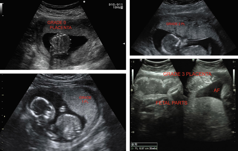
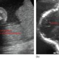
Figure 21.2 Illustrates varying grades of placental maturity in a normal fetus.
Related posts:
Stay updated, free articles. Join our Telegram channel

Full access? Get Clinical Tree


