Technical Considerations
In general, the pleura and chest wall are adequately evaluated with routine thoracic CT techniques. Contrast medium infusion is helpful in showing pleural thickening and in allowing its differentiation from pleural fluid. Soft-tissue window settings and bone windows are most suitable for evaluating pleural abnormalities, the chest wall, and the diaphragm.
It should be kept in mind that the diaphragm and posterior pleural space extend well below the lung bases, and scans inferior to the diaphragmatic domes must be obtained for their complete evaluation. A good general rule is that if ribs are visible, then the pleural space is being imaged. Scanning with the patient in the prone position may be of assistance in evaluating pleural diseases; free pleural effusions shift to the dependent portion of the pleural space when the patient is moved from the supine position to the prone or decubitus position, whereas loculated effusions or fibrosis show little or no change. Prone scans in patients with pleural effusion are most commonly obtained when CT is being used for chest tube placement
Pleura
Normal Anatomy
Because the lateral ribs are obliquely oriented, usually only a short segment of each rib is visible on a single CT slice. A more anterior rib represents one arising at a higher thoracic level than the rib posterior to it ( Fig. 7.1 ). Thus, for example, at any given level, the fifth rib is anterior to the sixth, the fourth is anterior to the fifth, and so on. At the level of the lung apex, the first rib can be identified by its anterior position and by its articulation with the manubrium immediately below the level of the clavicle.

In many patients a bony spur projects inferiorly from the undersurface of the first rib at its junction with the manubrium. In cross section, with a lung window, this bony spur can appear to be surrounded by lung and can mimic a lung nodule. This appearance is usually bilateral and symmetric, providing a clue as to its true nature. As would be expected, it appears calcified at tissue windows.
Costal Pleura
On thin-slice CT in normal individuals, a 1- to 2-mm-thick opaque stripe is commonly seen in the intercostal spaces, between adjacent rib segments ( Fig. 7.1 ). This stripe primarily represents the innermost intercostal muscle. In the paravertebral regions the innermost intercostal muscle is absent, and a much thinner line (or no line at all) is visible at the lung surface. The visceral and parietal pleura pass internal to the ribs and innermost intercostal muscles and are separated from them by a thin layer of extrapleural fat, but the pleural layers are not normally visible on CT.
Intrapulmonary Fissures
Intrapulmonary (interlobar) fissures are described and illustrated in Chapter 6 ( Fig. 6.1 ). Normal collections of fat extending into the inferior aspects of the major fissures at the diaphragmatic surface can simulate fissural pleural thickening or effusion. They appear low in attenuation at mediastinal window settings.
Inferior Pulmonary Ligament
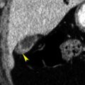
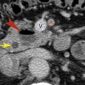
On each side below the inferior pulmonary vein, the parietal and visceral pleural layers join, forming a fold that extends inferiorly along the mediastinal surface of the lung and ends at the level of the diaphragm. This fold, the inferior pulmonary ligament , anchors the lower lobe. On CT images viewed at lung window settings, it appears as, or is related to, a small triangular opacity, 1 cm or less in size, with its apex pointing laterally into the lung and its base against the mediastinum. On each side, it usually lies adjacent to the esophagus ( Fig. 7.2 ). Pleural effusion or pneumothoraces can be limited and marginated by the inferior pulmonary ligament.

A similar opacity can be seen on the right, lateral to the inferior vena cava (and thus anterior to the inferior pulmonary ligament), and on the left arising lateral to the left ventricle, extending inferiorly to the diaphragm, then extending laterally for several centimeters along the diaphragmatic surface ( Fig. 7.2 ). These opacities represent the phrenic nerves and their pleural reflections. Their only significance is that they are commonly seen and just as commonly confusing.
Pleural Thickening and Look-alikes
Pleural thickening is visible on CT as a curvilinear soft-tissue stripe passing internal to the ribs and innermost intercostal muscles ( Fig. 7.1B ). A basic rule is that if the pleura is visible on CT, it is thickened. Thickened pleura enhances and is best seen after contrast medium infusion. Usually visible thickened pleura represents the parietal pleural layer.
In the presence of pleural thickening, the extrapleural fat layer is often thickened, and if this is the case, the visible pleural line is separated from the ribs and intercostal muscles by this fat layer ( Fig. 7.1B ). In the paravertebral regions or adjacent to the mediastinum, a distinct opaque stripe is visible in the presence of pleural thickening.
A small pleural effusion can mimic the appearance of pleural thickening; however, effusion is usually dependent on location and is crescentic. Thickened pleura can often be distinguished from small pleural fluid collections by contrast medium infusion; thickened pleura enhances, whereas fluid does not.
Normal extrapleural fat pads of a few millimeters in thickness can sometimes be seen internal to the ribs, particularly in the lower posterolateral thorax, and may not be easily distinguishable from pleural thickening or fluid with wide windows (1500 Hounsfield units [HU]). However, normal fat pads appear low in attenuation and are often symmetric, whereas pleural abnormalities generally are not.
The subcostalis muscles are sometimes visible posteriorly in the lower thorax as a 1- to 2-mm-thick stripe internal to one or more ribs. In contrast to pleural thickening, these muscles are smooth, uniform in thickness, and bilaterally symmetric.
Segments of intercostal veins are commonly visible in the paravertebral regions and can mimic focal pleural thickening. Continuity of these opacities with the azygos vein or hemiazygos vein can sometimes allow them to be identified correctly.
Pleural Effusion and Empyema
Diagnosis of Paradiaphragmatic Fluid Collections
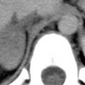
The visceral pleura covers the surface of the lung, and its inferior extent is defined by the inferior extent of the lung parenchyma. The parietal pleura is contiguous with the chest wall and diaphragm and extends well below the level of the bases of the lungs, into the costophrenic angles. Thus pleural fluid collections in the costophrenic angles can be seen below the lung base and can mimic collections of fluid in the peritoneal cavity.
The parallel curvilinear configuration of the pleural and peritoneal cavities at the level of the perihepatic and perisplenic recesses allows fluid in either cavity to appear as an arcuate or semilunar opacity displacing the liver or spleen away from the adjacent chest wall. The relation of the fluid collection to the ipsilateral diaphragmatic crus (see later discussion) helps to determine its location. Pleural fluid collections in the posterior costophrenic angle lie posterior to the diaphragm and cause lateral displacement of the crus. Peritoneal fluid collections are anterior to the diaphragm and lateral to the crus, displacing it medially. Fluid seen posterior to the liver is within the pleural space; the peritoneal space does not extend into this region (this is the “bare area” of the liver).
A large pleural effusion allows the lower lobe to float anteriorly and lose volume. The posterior edge of the lower lobe when surrounded by fluid both anteriorly and posteriorly can appear to represent the diaphragm (a pseudodiaphragm ), with pleural fluid posteriorly and ascites anteriorly ( Fig. 7.3 ). Sequential scans at more cephalad levels, however, generally allow the correct interpretation to be made. Typically, the arcuate opacity of the atelectatic lower lobe becomes thicker superiorly, is contiguous with the remainder of the lower lobe, and may contain air bronchograms.

Fissural Fluid
Large effusions often extend into the major fissures, displacing the lower lobes medially and posteriorly. A localized collection of pleural fluid in a major or minor fissure can have a confusing appearance on CT scans and can be misinterpreted as representing a parenchymal mass. However, careful analysis of contiguous images will usually confirm the relation of the mass to the plane of the fissure. The edges of the fluid collection may be seen to taper, conforming to the fissure and forming a “beak” ( Fig. 7.4 ).

Characterization of Pleural Effusion: Exudate or Transudate?
A pleural effusion is usually characterized by thoracentesis as being an exudate (a high-protein effusion associated with pleural disease) or a transudate (a low-protein effusion associated with alteration in systemic factors governing the formation of pleural fluid) ( Table 7.1 ). Exudates, being high in protein, tend to loculate, and tube drainage may be necessary. CT findings can help in diagnosing loculation and assessing the fluid collection.
| Exudates | Transudates |
|---|---|
| Parapneumonic effusion | Congestive heart failure |
| Empyema | Liver disease |
| Malignancy | Renal disease |
| Collagen vascular disease Pulmonary embolism | Overhydration Low serum protein level |
| Abdominal disease | |
| Hemothorax | |
| Chylothorax |
On CT a crescentic and dependent fluid collection is likely, but not always, free (a definite diagnosis of free pleural effusion requires demonstration of a shift in effusion in association with a shift in patient position). A crescentic effusion may be a transudate or an exudate ( Figs. 7.5 and 7.6 ). Lenticular (i.e., lens-shaped) fluid collections, and collections that are nondependent, are likely loculated, and thus very likely represent an exudate or an empyema ( Figs. 7.6 and 7.10 ). When one is describing an effusion on CT, its shape (crescentic or lenticular) and location (dependent or nondependent) should be indicated.


Most effusions appear to be near to water in attenuation, and measured CT numbers cannot be relied on to predict the specific gravity of the fluid or its nature (i.e., exudate or transudate). In patients with effusion the presence of pleural thickening on CT ( Figs. 7.6 and 7.7 ) predicts that the effusion is an exudate rather than a transudate, with a specificity of nearly 100%. By definition, the pleura is considered thickened on CT if it is visible; contrast enhancement helps greatly in making this diagnosis. Transudates are not associated with pleural thickening (except in the unusual case of a patient with preexisting pleural disease who develops an unrelated transudate).

The absence of pleural thickening on contrast-enhanced CT in a patient with pleural effusion is less helpful; in this situation the effusion can be an exudate or a transudate ( Fig. 7.7 ). Only about 50% to 60% of exudates are associated with visible pleural thickening on CT. However, the absence of pleural thickening on a contrast-enhanced scan makes empyema unlikely; empyema is almost always associated with parietal pleural thickening on contrast-enhanced CT.
Hemothorax
Hemothorax is defined as a pleural effusion having a hematocrit of at least 50% of the blood hematocrit. It may be traumatic or related to other causes of bleeding. Hemothorax may be dense (>50 HU) or may appear inhomogeneous, with some areas, particularly dependent regions, having an attenuation greater than that of the surrounding fluid. A fluid-fluid level (a hematocrit effect) or dependent clot may be seen with hemothorax ( Fig. 7.8 ).

Parapneumonic Effusion
Pleural fluid can accumulate in patients with pneumonia, even when the pleural space is uninfected. This is termed a simple parapneumonic effusion , and it results from increased permeability of the visceral pleura associated with the inflammation. The effusion is usually an exudate.
Parietal pleural thickening is present in about half of patients with parapneumonic effusion, whereas visceral pleural thickening is seen in one-fourth of patients. Because loculation is not usually present, the effusion is typically crescentic and dependent ( Fig. 7.9 ).

Empyema
Empyemas are often associated with pneumonia but may occur in the absence of lung infection. Empyema is diagnosed if pleural fluid contains infectious organisms on smear or culture.
Classically, an empyema is associated with the split pleura sign . This sign is said to be present when the thickened visceral and parietal pleural layers are split apart by and surround the infected fluid collection ( Figs. 7.7 and 7.10 ); the two pleural layers are generally similar in thickness. However, the split pleura sign is not always present in patients with empyema. Although empyema is nearly always associated with parietal pleural thickening on contrast-enhanced CT, visceral pleural thickening (and thus the split pleura sign) is present in only half of cases ( Fig. 7.6 ).

Empyemas can be free or loculated, and crescentic ( Fig. 7.6 ), rounded, elliptic, or lenticular ( Figs. 7.6, 7.7, and 7.10 ). Loculated effusions are typically, but not always, elliptic. Multiple loculations may be present ( Fig. 7.10B ).
In patients with a dependent and crescentic effusion associated with pleural thickening, simple parapneumonic effusion and empyema cannot be distinguished. However, loculation is not usually present in simple parapneumonic effusion and predicts empyema.
The presence of air within an empyema is almost always due to recent thoracentesis, but may also indicate bronchopleural fistula ( Fig. 7.11 ) or the presence of a gas-forming organism. In the absence of thoracentesis, this finding is usually an indication for tube drainage.

Extension of an empyema to involve the chest wall is termed empyema necessitatis. Two-thirds of cases result from tuberculosis, but other responsible organisms include Actinomyces and Nocardia. CT findings include low-attenuation fluid collections within the chest wall.
Differentiation of Empyema From Lung Abscess
Distinguishing empyema from lung abscess is sometimes important in patients who are clinically infected; empyema is often treated by tube drainage, whereas lung abscess generally is not. CT can be helpful in making this distinction, particularly when contrast medium is infused.
The outer edge of an empyema is sharply demarcated from adjacent lung, and on contrast-enhanced scans the empyema wall appears smooth and uniform in thickness. When a bronchopleural fistula is present and air is contained within the empyema cavity, or when air is introduced into the empyema during thoracentesis, its inner margin also appears smooth.
In contrast, lung abscesses are irregularly shaped and may contain multiple areas of low-attenuation necrosis or collections of air and fluid ( Fig 6.26 ). On contrast-enhanced scans the abscess wall is generally opacified relative to fluid in the abscess cavity ( Fig 6.26 ). The inner surfaces of an abscess may appear irregular and ragged, and their outer edges may be poorly defined or irregular because of adjacent pulmonary parenchymal consolidation.
At their point of contact with the chest wall, empyemas can show acute or obtuse angles ( Fig. 7.12 ), whereas abscesses typically have acute angles. Empyemas also tend to compress and displace the lung and vessels, acting as a space-occupying mass, whereas lung abscesses usually destroy the lung without displacing it.