Discussion
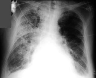
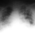
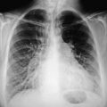
The portable film reveals infiltrates bilaterally. Note the basilar position of the infiltrates, quite characteristic of a patient who has been intubated for a long period of time and who has developed the classic appearance of aspiration pneumonia, specifically a nosocomial infection after prolonged intubation. The area of infection depends on the position of the patient. Classically, the pneumonia is in the basilar segments and in the most posterior segments of the lung.
A patient with chronic obstructive lung disease came to the emergency department with an elevated temperature. A portable film was obtained on admission to the ICU.
Discussion
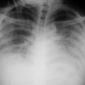
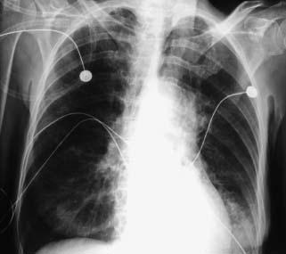
Note that this film is somewhat hyperaerated, but still one cannot see through the heart to visualize the vascularity of the lower lobes. In addition, one can see an area of infiltrate lateral to the left heart border, partially obliterating a portion of the left hemidiaphragm. The findings are all consistent with pneumonia in the left lower lobe.
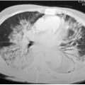
A patient who was in a brawl and was stabbed in the abdomen and chest was admitted several days earlier with a pneumothrax and perforated bowel. On postop day 6, he developed spiking temperatures and marked shortness of breath.
Discussion
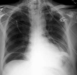
There is a significant amount of intra-abdominal air postoperatively, and a Heimlich tube with the pigtail opened up in the right chest.
Of interest, of course, is the left lower lobe. There is elevation in the left hemidiaphragm, with obliteration of the most medial portion of it. There is consolidation of the medial basal segments of the left lower lobe as well as areas of mediastinal emphysema.
This 88-year-old patient with high fever was admitted from a nursing home.
