Post-Primary Tuberculosis
Katherine R. Birchard
FINDINGS
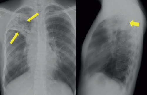
Figure 66A: Posteroanterior (PA) and lateral chest films show a heterogeneous opacity with bronchiectasis and cavities/cysts in the right upper lobe (yellow arrows), as well as elevation of minor fissure and superior retraction of right hilum.
DIFFERENTIAL DIAGNOSIS
Postprimary tuberculosis, lung cancer, sarcoid.
DIAGNOSIS
Postprimary tuberculosis (TB).
DISCUSSION
Parenchymal involvement in postprimary TB most commonly manifests as heterogeneous opacities in the apical and posterior segments of the upper lobes, and in the superior segment of the lower lobes, along with bronchiectasis, architectural distortion, calcifications, and residual cavities.1,2 Predilection of postprimary TB to involve the upper lobes is likely caused by the relatively higher oxygen tension and less robust lymphatic drainage. Because architectural distortion and calcifications may be present in both nonactive and active disease, determination of active TB infection based on radiographs alone is not possible, and must be confirmed by sputum culture,3 as was done in this patient.
Stay updated, free articles. Join our Telegram channel

Full access? Get Clinical Tree


