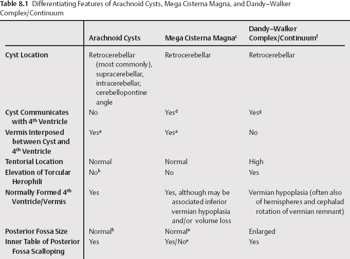
8 A variety of cystic lesions occur in the posterior fossa, with the most common being arachnoid cysts, Dandy-Walker complex/continuum, and mega cisterna magna (Table 8.1). It is often difficult to differentiate between these entities due to very subtle differences between them. Computed tomography (CT) cisternography can aid in the differentiation in certain cases, and some of these lesions can be correctly diagnosed on fetal magnetic resonance imaging (MRI). Ultrasound can also aid in the diagnosis. Thirteen to 30% of intracranial arachnoid cysts involve the posterior fossa and appear on CT as low-density, noncalcified, extraaxial masses that do not enhance. The higher incidence is reported in the pediatric literature, but in a study of patients of mixed ages with adult predominance, 13% of 93 arachnoid cysts occurred in the posterior fossa. The spectrum of anomalies defined by varying degrees of vermis hypoplasia (with cephalad rotation of the vermian remnant), retrocerebellar cyst that communicates with the 4th ventricle, and enlarged posterior fossa with elevation of the torcular Herophili has been variously termed Dandy-Walker malformation, Dandy-Walker variant, Dandy-Walker continuum, Dandy-Walker spectrum, and Dandy-Walker complex.
Posterior Fossa Cysts and Cerebellar Malformations
Arachnoid Cysts
Dandy-Walker Continuum
![]()
Stay updated, free articles. Join our Telegram channel

Full access? Get Clinical Tree


