Posterior Hip Dislocation with Intra-Articular Fragment
Andrew F. Barnes
Daniel B. Nissman
CLINICAL HISTORY
39-year-old male presents with right hip pain following a front-end motor vehicle collision.
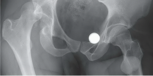 FIGURE 92A |
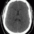
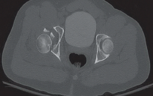 FIGURE 92B |
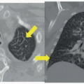
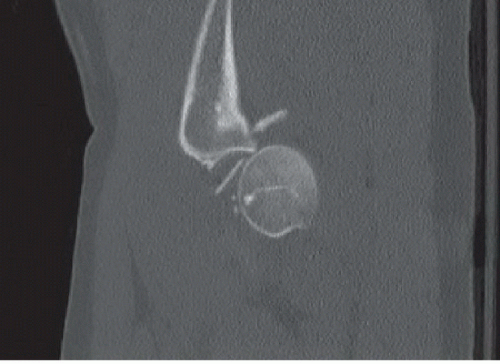 FIGURE 92C |
FINDINGS
Supine AP radiograph of the pelvis (Fig. 92A) demonstrates a dislocated right hip, with marked superior displacement of the femoral head; the right femur is internally rotated relative to the left. There is a vertically oriented fracture of the posterior acetabular wall. Axial CT image of the pelvis at the level of the right femoral head following attempted reduction (Fig. 92B) demonstrates partial reduction with persistent posterior and superior subluxation. Two large intra-articular osseous bodies are present. Sagittal CT image through the right hip (Fig. 92C) demonstrates posterior subluxation of the femoral head and both intra- and extraarticular bone fragments.
DIFFERENTIAL DIAGNOSIS
Hip dislocation (anterior or posterior) with or without acetabular fracture or femoral head fracture.
Related posts:


Stay updated, free articles. Join our Telegram channel

Full access? Get Clinical Tree


