Posterior Mediastinal Hematoma
Katherine R. Birchard
CLINICAL HISTORY
36-year-old male after a motor vehicle accident.
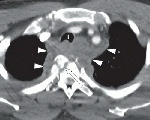 FIGURE 62A |
FINDINGS FIGURE 62A:
Axialcontrast-enhanced CT image of the chest just below thoracic inlet shows comminuted T4 vertebral body fracture (arrow) and abnormal high-density soft tissue (arrowheads) in the paraspinous region, posterior mediastinum, and extending into the middle mediastinum, displacing the trachea (t) and great vessels anteriorly.
DIFFERENTIAL DIAGNOSIS
Posterior mediastinal hematoma, neurogenic tumor, osteomyelitis.
DIAGNOSIS
Related posts:


Stay updated, free articles. Join our Telegram channel

Full access? Get Clinical Tree


