Magnetic resonance imaging (MRI) and MR arthrography have proven invaluable for managing the postoperative shoulder, particularly in relation to the rotator cuff and labrum. MRI has proven to be an accurate imaging technique for differentiating expected findings versus complications in the postoperative setting. The transition from metallic hardware to bioabsorbable suture anchors used in orthopedic surgery has rendered less metallic susceptibility artifact over the years, allowing more accurate interpretation of MR images. This article gives a pictorial review of various expected postoperative findings in the shoulder and complications related to repair of the rotator cuff and labrum.
Magnetic resonance imaging (MRI) and MR arthrography have proven invaluable for the management of the postoperative shoulder, particularly in relation to the rotator cuff and labrum. MRI has proven to be an accurate imaging technique for the differentiation of expected findings versus complications in the postoperative setting. The transition from metallic hardware to bioabsorbable suture anchors used in orthopedic surgery has rendered less metallic susceptibility artifact over the years, allowing a more accurate interpretation of MR images. This article gives a pictorial review of various expected postoperative findings in the shoulder and complications related to repair of the rotator cuff and labrum.
Technical considerations
The preferred imaging methods for evaluating the postoperative shoulder include MR arthrography, conventional MRI, and sonography (depending on the expertise of the interpreting radiologist). When bioabsorbable anchors and other nonmetallic devices have been used, shoulder MR arthrography or conventional MRI are acquired in a similar fashion as in the preoperative patient, as outlined in the American College of Radiology-Society of Skeletal Radiology (ACR-SSR) Practice Guideline for the Performance and interpretation of Magnetic Resonance Imaging (MRI) of the Shoulder. Metallic suture anchors and shoulder arthroplasties do not pose an MRI safety risk for patients, but techniques should be implemented to decrease metallic artifact. These include increasing the bandwidth, increasing the of echo train, using a small field of view, increasing the matrix, using fast STIR (short tau inversion recovery) sequences as opposed to fat suppressed sequences, and avoiding gradient echo imaging.
Although this article is limited to the use of MRI for evaluating the postoperative shoulder, computed tomography (CT) arthrography is regarded as superior to MRI or MR arthrography in the evaluation of the rotator cuff in the setting of a previous shoulder arthroplasty. However, CT arthrography is viewed as a second-line procedure for evaluating shoulders with suspected instability or labral disorders when MRI is not available or contraindicated. Contraindications to MRI include the presence of non-MRI compatible intracranial aneurysm clips, pacemakers, implanted cardioverter defibrillators, intraorbital metal, and a variety of electronic and magnetically activated implants, devices, stents, and coils.
Expected postoperative findings
Rotator Cuff Repair
Arthroscopic repair of rotator cuff tears generally yields excellent pain relief and improvement in the ability to perform daily activities, despite structural failure demonstrated on MRI. Early repair of traumatic rotator cuff tears provides better results in terms of shoulder function, in comparison with delayed tendon repair. The factors affecting tendon healing are patient age, the size and extent of the tear, and the presence of fatty degeneration of the rotator cuff muscle. Although functional status tends to improve with time after 6 months, the structural status of repaired cuffs remains unchanged and can be well evaluated with MRI. Repaired rotator cuff tendons will have a variable appearance on MRI, with temporal evolution on serial imaging. Within 3 months of operating, tendons appear most disorganized compared with native tendons, and intermediate signal intensity within the surgical bed will correspond to granulation tissue. With time, the development of fibrotic tissue will yield areas of low signal intensity on all sequences. Of note, only 10% of repaired tendons demonstrate normal signal intensity on MRI.
Partial-Thickness Rotator Cuff Tendon Tear Repair
If partial-thickness rotator cuff tears are treated surgically, they may be debrided, repaired with a transtendon technique, or repaired after tear completion. When tears are bursal sided and there are morphologic changes in the coracoacromial arch, a subacromial decompression may also be performed. Repair of high-grade articular sided tears may also be accompanied by anterior acromioplasty. When tears are debrided, they typically have a defect that is longer than deep relative to the long axis of the tendon on MRI ( Fig. 1 ). If the converse is true, then a recurrent tear is likely. In a study of 48 patients treated for high-grade partial-thickness rotator cuff tears, repair after conversion to a full-thickness tear showed less postoperative morbidity compared with those treated with transtendon technique; however, recurrent tears developed in 8% of those repaired tendons after tear completion.
The transosseous equivalent repair technique is designed for small to medium U-shaped tears and for iatrogenically completed partial articular supraspinatus tendon avulsions of moderate to large size. The use of selective knot placement allows the surgeon to convert a linear construct into a V configuration, which will optimize repair strength and allow earlier rehabilitation due to increased footprint contact dimensions and less repair gap.
Full-Thickness Rotator Cuff Tendon Tear Repair
Direct suture repair
Rotator cuff tendon tears involving the myotendinous junction or critical zone may be treated with direct suture repair. There is often a slight difference in tendon caliber at the reattachment ( Fig. 2 ), and, as with any repaired tendon, only 10% of repaired tendons will maintain the normal, native low signal intensity.
Single Row Technique
Most small full-thickness rotator cuff repairs are performed by using the tendon-to-bone repair technique, and various surgical tacks and suture material are available on the market. Assessment of the integrity of repaired rotator cuff tendon on postoperative MRI must address both the suture anchors and the tendon. Osteolysis around bioabsorbable suture anchors is an expected reaction ( Fig. 3 ), and this is caused by mechanical forces or focal necrosis resulting from drilling. These osseous lucencies may double in size at 6 months, but should eventually stabilize and become replaced with bone at 2 years.
Two-tendon tears of the rotator cuff can heal at a high rate with the use of transosseous-equivalent (TOE) suture bridge repair technique. In a study by Sethi and colleagues, 83% of the repairs demonstrated intact rotator cuff repairs at a mean of 16 months postoperatively. Larger tears (3.5 vs 2.8 cm) were associated with failure ( P = .01), as was more advanced fatty infiltration (Goutallier 1.3 vs 0.3, P = .01). Furthermore, the modified transosseous equivalent procedure, otherwise known as surface holding repair with transosseous sutures, has also been used for massive rotator cuff tears involving at least 2 tendons, with a 92% continuous rotator cuff on postoperative MRI scans.
Of note, asymptomatic patients may have signal changes suggestive of tendinopathy and have clinically silent partial and complete rotator cuff tears. In addition, mild bone marrow edema-like signal changes may be present up to 5 years after surgery in asymptomatic patients who have undergone rotator cuff repair. Patients can have marked improvement in symptoms without a watertight seal. Suspension bridge repair of massive rotator cuff tears can be effective, and complete coverage is not essential to convert debilitating tears into functional cuff tears.
Double-Row Technique
Double-row suture anchor repair is done in conjunction with creation of an implantation trough at the junction of the humeral head and greater tuberosity, which allows optimal apposition of tendon to bone ( Fig. 4 ). Intact tendons will have heterogeneous signal on postoperative MRI in the majority of cases ( Fig. 5 ). Ma and colleagues compared the MR arthrographic follow-up of 53 repaired rotator cuff tears that were performed with either single- or double-row technique. Although there was better shoulder strength demonstrated in patients who underwent double-row repair for larger size tears, there was no significant difference in postoperative structural integrity between the 2 groups at 6-month and 2-year follow-up with MR arthrography, regardless of tear size. Koh and colleagues did not find a statistically significant difference between the retear rates of arthroscopic single-row and double-row suture anchor repair in rotator cuff tears measuring between 2 and 4 cm. The short-term results of the clinical outcomes and structural integrity of TOE double-row rotator cuff repair (the suture–bridge technique) have results that compare favorably with those reported for other double-row suture anchor techniques employed in rotator cuff repairs. The geometry of the TOE mattress suture configuration construct compresses the tendon, optimizing tendon-to-tuberosity contact dimensions in an attempt to restore the native footprint, while providing strength sufficient to withstand immediate postoperative rehabilitation.
Patch Grafting
Irreparable massive rotator cuff tears can be repaired using arthroscopic rotator cuff reconstruction with a variety of patch grafts, although there is limited postoperative imaging available to date in the literature. Patch grafts may be composed of allogenic freeze-dried tissues, artificial synthetic grafts, porcine small intestine submucosa, or autografts (such as the long head of the biceps). Of note, all of the allografts have the potential to incite an aseptic inflammatory or foreign body reaction, which is evidenced by edema-like signal changes throughout the soft tissues and potentially the osseous structures on MRI.
Certain centers are now using polycarbonate polyurethane patches secured in a 6-point fixation construct to augment rotator cuff repairs. In a study of 10 patients at 1 year follow-up, Encaalada-Diaz and colleages found a 10% retear rate,on small and medium sized tears, and no visible subacromial adhesions on follow-up MRI using these nonresorbable scaffolds for permanent structural support. Since these constructs are so thin and are adherent to the repaired tendons, they are not well visualized on postoperative MRI. GraftJacket (Wright Medical Group, Incorporated, Arlington, TN, USA) is 1 type of bridging allograft that is composed of acellular human dermal matrix. This construct is implanted along the greater tuberosity and provides added support to the reattached tendons.
Biceps Tenotomy/Tenodesis
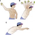
Tenotomy or tenodesis of the long head of the biceps is performed if there are irreversible structural changes in the tendon, significant atrophy or hypertrophy, partial tearing greater than 25% of the width of the tendon, subluxation of tendon from the groove, or if there are certain disorders of the biceps origin. Tenodesis remains the preferred treatment for younger patients with biceps dysfunction ( Fig. 6 ).
Labral Tear Repair
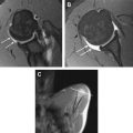
Direct anatomic repair of labral tears usually occurs by suturing the anterior labrum and joint capsule, and the anterior band of the inferior band of the inferior glenohumeral ligament (IGHL) to the glenoid rim. With MRI or MR arthrography, there should be no separation of the labrocapsular complex and glenoid margin in intact labral repairs ( Figs. 7–9 ). Susceptibility artifact from metallic fixation should be minimized with MRI metal reduction techniques. Capsular thickening with an irregular nodular contour is an expected postoperative finding. Granulation tissue within the repaired labral tear may prevent joint fluid or injected contrast from outlining a persistent defect, resulting in false-negative MR examinations. Comparison with prior MRI studies is of utmost importance to evaluate for retear. The overall accuracy of MR arthrography for detecting labral tears after prior instability repair varies in the literature, but is generally greater than 90%.
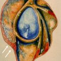
Paralabral cysts can develop through labral tears and extend along the spinoglenoid or suprascapular notches. These are known as spinoglenoid notch or suprascapular notch ganglia. Gains in external rotation strength have been shown in patients who are treated with cyst decompression and superior labrum anterior and posterior (SLAP) repair, as opposed to SLAP repair alone ( Fig. 10 ).
Recurrent anterior shoulder dislocation typically results in combined Bankart and Hill-Sachs lesions, although concomitant SLAP lesions may also be present. Arthroscopic Bankart repair after recurrent anterior shoulder dislocation may be performed in conjunction with the remplissage technique for treatment of instability with engaging Hill-Sachs lesions. This technique transfers the posterior capsule and infraspinatus tendon into the Hill-Sachs lesion to prevent engagement of the lesion on the glenoid rim. MRI will show corresponding findings of reattachment of the posterior structures into the defect, along with the metallic or bioabsorbable anchor embedded in the trough.
Other Postoperative Conditions
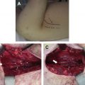
MRI may be helpful to assess the integrity of soft tissue reconstructions, such as reconstructed coracoclavicular ligaments after grade 3 or higher acromioclavicular (AC) joint separation ( Fig. 11 ). MRI allows for direct visualization of the cable components and anatomic alignment of the AC joint, confirming that the construct is intact.
MRI is mainly used preoperative planning for shoulder arthroplasty to detect any rotator cuff tears. MRI after shoulder arthroplasty can result in various degrees of susceptibility artifact, depending on the type of metallic construct ( Fig. 12 ). MRI may be a helpful adjunct to radiographs and CT in select cases to determine the extent of particle disease in the adjacent bones and soft tissues.
Expected postoperative findings
Rotator Cuff Repair
Arthroscopic repair of rotator cuff tears generally yields excellent pain relief and improvement in the ability to perform daily activities, despite structural failure demonstrated on MRI. Early repair of traumatic rotator cuff tears provides better results in terms of shoulder function, in comparison with delayed tendon repair. The factors affecting tendon healing are patient age, the size and extent of the tear, and the presence of fatty degeneration of the rotator cuff muscle. Although functional status tends to improve with time after 6 months, the structural status of repaired cuffs remains unchanged and can be well evaluated with MRI. Repaired rotator cuff tendons will have a variable appearance on MRI, with temporal evolution on serial imaging. Within 3 months of operating, tendons appear most disorganized compared with native tendons, and intermediate signal intensity within the surgical bed will correspond to granulation tissue. With time, the development of fibrotic tissue will yield areas of low signal intensity on all sequences. Of note, only 10% of repaired tendons demonstrate normal signal intensity on MRI.
Partial-Thickness Rotator Cuff Tendon Tear Repair
If partial-thickness rotator cuff tears are treated surgically, they may be debrided, repaired with a transtendon technique, or repaired after tear completion. When tears are bursal sided and there are morphologic changes in the coracoacromial arch, a subacromial decompression may also be performed. Repair of high-grade articular sided tears may also be accompanied by anterior acromioplasty. When tears are debrided, they typically have a defect that is longer than deep relative to the long axis of the tendon on MRI ( Fig. 1 ). If the converse is true, then a recurrent tear is likely. In a study of 48 patients treated for high-grade partial-thickness rotator cuff tears, repair after conversion to a full-thickness tear showed less postoperative morbidity compared with those treated with transtendon technique; however, recurrent tears developed in 8% of those repaired tendons after tear completion.
Related posts:
 Magnetic Resonance Imaging of Rotator Cuff Disease and External Impingement
 Internal Impingement Syndromes
 The Throwing Shoulder: the Orthopedist Perspective
Magnetic Resonance Imaging of the Pediatric Shoulder
The Throwing Shoulder: the Orthopedist Perspective
Magnetic Resonance Imaging of the Pediatric Shoulder
 Entrapment Neuropathies of the Shoulder
 Anatomic Variants and Pitfalls of the Labrum, Glenoid Cartilage, and Glenohumeral Ligaments
Stay updated, free articles. Join our Telegram channel

Full access? Get Clinical Tree


