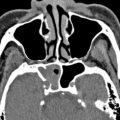
Endoscopic sinus surgery is a minimally invasive option for the treatment of several nonneoplastic indications, particularly for medically refractory sinusitis and polyposis. Numerous interventions can be performed through endoscopic sinus surgery, many of which may be performed together during the same procedure. There are also a variety of complications that can result from endoscopic sinus surgery. Radiological imaging plays an important role in the evaluation of patients after endoscopic sinus surgery. Thus, it is important to be familiar with the expected and complicated imaging findings associated with endoscopic sinus surgery, which are reviewed in this article.
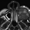
Complications resulting from endoscopic sinus surgery are summarized and selected examples of diagnostic imaging are presented.
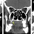
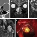
Complications resulting from endoscopic sinus surgery are summarized and selected examples of diagnostic imaging are presented.
Ultimately the decision to obtain diagnostic imaging following endoscopic sinus surgery depends on the specific clinical situation. The role of radiologists may be to guide the referring physicians toward selecting the most appropriate imaging modality for the particular clinical question ( Table 3 ). The goal of radiologists is also to provide a systematic description of the apparent postoperative findings, both expected and unexpected, with attention to the various conditions reviewed in this article.
| Clinical Question | Suggested Imaging Modalities |
|---|---|
| Where is the site of CSF leak? | CT |
| Is there a cephalocele? | Skull base MR imaging |
| What is the source of the bleeding? | Catheter angiography or CTA |
| Is there intracranial infection? | Brain MR imaging with contrast |
| Why are there vision changes? | Orbit CT and/or MR imaging |
| Why is there recurrent sinusitis? | Sinus CT |
| Why is there nasal obstruction? | Sinus CT |
References
- 1. Ginat D.T., and Cunnane M.E.: Imaging the paranasal sinuses and nasal cavity after surgery. In Ginat D.T., and Westesson P.L. (eds): Atlas of postsurgical neuroradiology. Berlin: Springer, 2012. pp. 75-120
- 2. Bhatti M.T.: Neuro-ophthalmic complications of endoscopic sinus surgery. Curr Opin Ophthalmol 2007; 18: pp. 450-458
- 3. Bhatti M.T., Schmalfuss I.M., and Mancuso A.A.: Orbital complications of functional endoscopic sinus surgery: MR and CT findings. Clin Radiol 2005; 60: pp. 894-904
- 4. Zeifer B.: Sinusitis: postoperative changes and surgical complications. Semin Ultrasound CT MR 2002; 23: pp. 475-491
- 5. Tan V.E., and Sethi D.S.: Gossypiboma: an unusual intracranial complication of endoscopic sinus surgery. Laryngoscope 2011; 121: pp. 879-881
- 6. Krings J.G., Kallogjeri D., Wineland A., et al: Complications of primary and revision functional endoscopic sinus surgery for chronic rhinosinusitis. Laryngoscope 2014; 124: pp. 838-845
- 7. Valdes C.J., Bogado M., and Samaha M.: Causes of failure in endoscopic frontal sinus surgery in chronic rhinosinusitis patients. Int Forum Allergy Rhinol 2014; 4: pp. 502-506
- 8. May M., Levine H.L., Mester S.J., et al: Complications of endoscopic sinus surgery: analysis of 2108 patients–incidence and prevention. Laryngoscope 1994; 104: pp. 1080-1083
- 9. Yang B.T., Liu Y.J., Wang Y.Z., et al: CT and MR imaging findings of periorbital lipogranuloma developing after endoscopic sinus surgery. AJNR Am J Neuroradiol 2012; 33: pp. 2140-2143
- 10. Available at: http://www.guideline.gov/content.aspx?id=37936&search=Chronic+sinusitis. Accessed February 1, 2015.
- 11. Cunnane M.E., Platt M., Caruso P.A., et al: Medialization of the lamina papyracea after endoscopic ethmoidectomy: comparison of preprocedure and postprocedure computed tomographic scans. J Comput Assist Tomogr 2009; 33: pp. 79-81
- 12. Albu S., Gocea A., and Necula S.: Simultaneous inferior and middle meatus antrostomies in the treatment of the severely diseased maxillary sinus. Am J Rhinol Allergy 2011; 25: pp. e80-e85
- 13. Draf W., Weber R., Keerl R., et al: Aspects of frontal sinus surgery. Part I: Endonasal frontal sinus drainage for inflammatory sinus disease. HNO 1995; 43: pp. 352-357
- 14. Rains B.M.: Frontal sinus stenting. Otolaryngol Clin North Am 2001; 34: pp. 101-110
- 15. Moeller C.W., and Welch K.C.: Prevention and management of complications in sphenoidotomy. Otolaryngol Clin North Am 2010; 43: pp. 839-854
- 16. Kieff D.A., and Busaba N.: Treatment of isolated sphenoid sinus inflammatory disease by endoscopic sphenoidotomy without ethmoidectomy. Laryngoscope 2002; 112: pp. 2186-2188
- 17. Donald P.J.: Sphenoid marsupialization for chronic sphenoidal sinusitis. Laryngoscope 2000; 110: pp. 1349-1352
- 18. Leight W.D., and Leopold D.A.: Sphenoid “drill-out” for chronic sphenoid rhinosinusitis. Int Forum Allergy Rhinol 2011; 1: pp. 64-69
- 19. Levine H., and Rabago D.: Balloon sinuplasty: a minimally invasive option for patients with chronic rhinosinusitis. Postgrad Med 2011; 123: pp. 112-118
- 20. Taghi A.S., Khalil S.S., Mace A.D., et al: Balloon sinuplasty: balloon-catheter dilation of paranasal sinus ostia for chronic rhinosinusitis. Expert Rev Med Devices 2009; 6: pp. 377-382
- 21. Barrow E.M., and DelGaudio J.M.: In-office drainage of sinus mucoceles: an alternative to operating-room drainage. Laryngoscope 2015; 125: pp. 1043-1047
- 22. Sautter N.B., Citardi M.J., Perry J., et al: Paranasal sinus mucoceles with skull-base and/or orbital erosion: is the endoscopic approach sufficient? Otolaryngol Head Neck Surg 2008; 139: pp. 570-574
- 23. Khong J.J., Malhotra R., Wormald P.J., et al: Endoscopic sinus surgery for paranasal sinus mucocoele with orbital involvement. Eye (Lond) 2004; 18: pp. 877-881
- 24. Alobid I., Bernal M., Calvo C., et al: Treatment of rhinocerebral mucormycosis by combination of endoscopic sinus debridement and amphotericin B. Am J Rhinol 2001; 15: pp. 327-331
- 25. Schatz C.J., and Ginat D.T.: Imaging features of rhinoplasty. AJNR Am J Neuroradiol 2014; 35: pp. 216-222
- 26. Guyuron B., and Vaughan C.: Evaluation of stents following septoplasty. Aesthetic Plast Surg 1995; 19: pp. 75-77
- 27. Egan K.K., and Kim D.W.: A novel intranasal stent for functional rhinoplasty and nostril stenosis. Laryngoscope 2005; 115: pp. 903-909
- 28. Bäck L.J., Hytönen M.L., Malmberg H.O., et al: Submucosal bipolar radiofrequency thermal ablation of inferior turbinates: a long-term follow-up with subjective and objective assessment. Laryngoscope 2002; 112: pp. 1806-1812
- 29. Demir U., Durgut O., Saraydaroglu G., et al: Efficacy of radiofrequency turbinate reduction: evaluation by computed tomography and acoustic rhinometry. J Otolaryngol Head Neck Surg 2012; 41: pp. 274-281
- 30. Metson R., and Pletcher S.D.: Endoscopic orbital and optic nerve decompression. Otolaryngol Clin North Am 2006; 39: pp. 551-561
- 31. Sciarretta V., Pasquini E., Tesei F., et al: Endoscopic sinus surgery for the treatment of maxillary sinus atelectasis and silent sinus syndrome. J Otolaryngol 2006; 35: pp. 60-64
- 32. La Fata V., McLean N., Wise S.K., et al: CSF leaks: correlation of high-resolution CT and multiplanar reformations with intraoperative endoscopic findings. AJNR Am J Neuroradiol 2008; 29: pp. 536-541
- 33. DelGaudio J.M., Baugnon K.L., Wise S.K., et al: Magnetic resonance cisternogram with intrathecal gadolinium with delayed imaging for difficult to diagnose cerebrospinal fluid leaks of anterior skull base. Int Forum Allergy Rhinol 2015; 5: pp. 333-338
- 34. Weidenbecher M., Huk W.J., and Iro H.: Internal carotid artery injury during functional endoscopic sinus surgery and its management. Eur Arch Otorhinolaryngol 2005; 262: pp. 640-645
- 35. Hudgins P.A., Browning D.G., Gallups J., et al: Endoscopic paranasal sinus surgery: radiographic evaluation of severe complications. AJNR Am J Neuroradiol 1992; 13: pp. 1161-1167
- 36. Bolger W.E., Parsons D.S., Mair E.A., et al: Lacrimal drainage system injury in functional endoscopic sinus surgery. Incidence, analysis, and prevention. Arch Otolaryngol Head Neck Surg 1992; 118: pp. 1179-1184
- 37. Chung S.K., Cho D.Y., and Dhong H.J.: Computed tomogram findings of mucous recirculation between the natural and accessory ostia of the maxillary sinus. Am J Rhinol 2002; 16: pp. 265-268
- 38. Albu S., Tomescu E., Mexca Z., et al: Recurrence rates in endonasal surgery for polyposis. Acta Otorhinolaryngol Belg 2004; 58: pp. 79-86
- 39. Larsen K., and Tos M.: A long-term follow-up study of nasal polyp patients after simple polypectomies. Eur Arch Otorhinolaryngol 1997; 254: pp. S85-S88
- 40. Topal O., Celik S.B., Erbek S., et al: Risk of nasal septal perforation following septoplasty in patients with allergic rhinitis. Eur Arch Otorhinolaryngol 2011; 268: pp. 231-233
- 41. Mullace M., Gorini E., Sbrocca M., et al: Management of nasal septal perforation using silicone nasal septal button. Acta Otorhinolaryngol Ital 2006; 26: pp. 216-218
- 42. Sozansky J., and Houser S.M.: Pathophysiology of empty nose syndrome. Laryngoscope 2014; 125: pp. 70-74
- 43. Coste A., Dessi P., and Serrano E.: Empty nose syndrome. Eur Ann Otorhinolaryngol Head Neck Dis 2012; 129: pp. 93-97
Related posts:

Stay updated, free articles. Join our Telegram channel

Full access? Get Clinical Tree


