Glioma is considered the most common type of primary central nervous system (CNS) tumor. Imaging is crucial for diagnosis, characterization, grading, and therapeutic planning of CNS gliomas. Along with a brief description of conventional computed tomography and magnetic resonance imaging techniques, this article reviews the ever-developing role of modern imaging techniques in preoperative management of CNS gliomas. It discusses current clinical applications, promising features, and limitations of each imaging method.
Key points
- •
Despite their limited role, conventional computed tomography and magnetic resonance (MR) imaging are traditionally considered the primary techniques for characterization of central nervous system (CNS) glioma; however, advanced imaging methods have evolved the imaging of glioma.
- •
Various modern imaging techniques, such as diffusion-weighted imaging, perfusion imaging, MR spectroscopy, and susceptibility-weighted imaging, might be used to assess presurgical grading, prognosis, and biopsy planning of CNS gliomas.
- •
The use of functional MR imaging and diffusion tensor imaging to delineate the relation of the tumor to important anatomic and functional areas has been promising. These techniques are shown to be useful in decreasing postoperative disability and allowing maximum tumor resection. Both techniques are subject to various limitations and need further technical improvements.
- •
PET is valuable for evaluation of the various aspects of the cellular metabolism of gliomas, which can potentially aid in surgical planning of gliomas and predicting their prognosis.
Introduction
Glioma, the most common primary brain tumor, refers to all the tumors originating from glial cells. Gliomas constitute approximately 27% of all primary central nervous system (CNS) tumors and 80% of malignant tumors. Glioma includes all the neoplasms arising from astrocytic, ependymal, and oligodendroglial cells or choroid plexus. Astrocytomas (originating from astrocytes) can be either circumscribed or diffuse. Circumscribed astrocytomas include pilocytic astrocytoma, pleomorphic xanthoastrocytoma, and subependymal giant cell astrocytoma. Diffuse astrocytomas consist of low-grade fibrillary astrocytoma, anaplastic astrocytoma, gliomatosis cerebri, and glioblastoma multiforme (GBM). Based on the World Health Organization (WHO) guidelines, gliomas are classified into 4 grades ( Table 1 ). Mortality increases in higher-grade tumors but, depending on the anatomic location, and the tendency for local infiltration and later malignant transformation, even grade I tumors can be fatal.
| Tumor Origin | Grade 1 | Grade 2 | Grade 3 | Grade 4 |
|---|---|---|---|---|
| Astrocytic |
|
| Anaplastic astrocytoma |
|
| Oligodendroglial | — |
| Anaplastic oligodendroglioma/oligoastrocytoma | — |
| Ependymal | Subependymoma | Ependymoma | Anaplastic ependymoma | — |
| Choroid plexus | Choroid plexus papilloma | Atypical choroid plexus papilloma | Choroid plexus carcinoma | — |
| Others | Angiocentric glioma | Chordoid glioma | — | — |
Imaging plays an important role in the diagnosis and management of gliomas. In the past, computed tomography (CT) scan and conventional magnetic resonance (MR) imaging techniques were used for detection and characterization of gliomas. With recent advances in brain tumor imaging, preoperative assessment of glioma has progressed. Modern imaging techniques evaluate all aspects of initial diagnosis, including the extent of disease, grading, surgical planning, and prognosis. The gold standard in the diagnosis and grading of gliomas remains histopathology. However, in daily practice, because the sampling is difficult and subject to possible morbidities, the therapeutic plan of many individuals with CNS glioma is performed based on the imaging features. The use of preoperative functional and anatomic imaging methods has evolved the therapeutic plans, resulting in safer surgical approach, increased survival, and decreased postoperative functional disability.
This article discusses the role of preoperative routine conventional imaging in individuals with CNS gliomas, along with more detailed description of specific modern imaging methods.
Conventional imaging techniques
CT might be used as the primary modality for identification of the brain mass, but its role in the characterization of primary CNS neoplasms and differentiation from nonneoplastic conditions is limited. The appearance of astrocytomas on CT scan varies from usually homogeneous in low-grade gliomas to heterogeneous in higher grades. CT scan is usually considered superior to conventional MR imaging for demonstration of intratumoral calcification, most frequently seen in oligodendrogliomas. Conventional MR imaging is the mainstay of CNS glioma imaging. However, it is not accurate enough for differentiation of tumor subtypes and estimation of tumor grade. Exceptions are juvenile pilocytic astrocytoma, subependymal giant cell astrocytoma, and pleomorphic xanthoastrocytoma, which show characteristic features on conventional MR imaging.
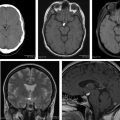
Overall, low-grade gliomas are hypointense on T1 and hyperintense on T2 with absent or minimal mass effect. As the tumor progresses to higher grades, it becomes more heterogeneous with ill-defined and irregular borders, apparent peripheral edema, and mass effect ( Fig. 1 A, B ) Hemorrhage may contribute to the heterogeneity of these tumors. Central coagulative necrosis is the hallmark of grade 4 glioma. Intravenous gadolinium-based agents increase the accuracy of MR imaging. In general, the presence of contrast enhancement is associated with higher-grade gliomas ( Fig. 1 C). The peripheral, irregular, and nodular enhancement is characteristic for GBM, whereas anaplastic astrocytoma shows patchy or no enhancement. It is notable that several subtypes of low-grade gliomas show contrast enhancement; the most important is the mural nodule of juvenile pilocytic astrocytoma and pleomorphic xanthoastrocytoma. In addition, nonenhancing gliomas are reported to be high grade in about one-third of cases. In high-grade gliomas, infiltration of tumoral cells into surrounding edema beyond the margin of enhanced area is described as infiltrative edema. Conventional MR imaging techniques are unable to show the areas of infiltrative edema. Altogether, conventional CT and MR imaging are not accurate modalities to characterize low-grade and high-grade gliomas.
Modern imaging techniques
Magnetic Resonance Perfusion Imaging
Neoangiogenesis is vital for glial tumor growth. Traditionally, the tumor vascularity is evaluated with contrast-enhanced CT or MR imaging, although the pattern of enhancement in gliomas does not allow accurate estimation of tumor vascular density, because it depends on the integrity of the blood-brain barrier, the amount of the contrast in the intra-arterial compartment, diffusion of the extravasated contrast to the adjacent tissues, and reabsorption of the contrast. Dynamic CT and MR perfusion imaging are more accurate noninvasive methods for assessment of tumor vascularity. MR perfusion can be performed either with or without intravenous contrast. The favored techniques of contrast-enhanced MR perfusion include T2 ∗ -weighted dynamic susceptibility contrast-enhanced (DSC) and T1-weighted dynamic contrast-enhanced (DCE). These techniques allow calculation of the cerebral blood volume (CBV) and endothelial transfer coefficient (K trans ) respectively. DSC is the most common perfusion technique used in preoperative glioma imaging for a range of applications, including pretherapeutic glioma grading, differentiating high-grade glioma from CNS lymphoma or solitary brain metastases, and predicting prognosis. The most common perfusion parameter to assess glioma angiogenesis is the relative CBV (rCBV). rCBV has reportedly shown the ability to differentiate low-grade from high-grade glioma, but it can be misleading in oligodendrogliomas (because of the so-called chicken-wire capillary architecture) and pilocytic astrocytomas. Overall, low-grade gliomas show normal/minimally increased rCBV. As the tumor transforms to higher grades, rCBV value increases, with GBMs showing markedly increased rCBV ( Figs. 1 F, 2 , and 4 D ). Moreover, by showing the most malignant part of a CNS tumor, rCBV maps can be used to guide biopsy.
The second MR perfusion technique with intravenous contrast agent is DCE. The advantage of DCE is the superior spatial resolution, less susceptibility artifact, and three-dimensional acquisitions. K trans is the main indicator of capillary permeability and neoangiogenesis in DCE. Similar to rCBV, there is evidence that K trans is an effective way to distinguish high-grade from low-grade glioma. DCE is limited because institutions use different sorts of postprocessing algorithms to calculate K trans ; this needs to be standardized for future studies.
The third MR perfusion technique, arterial spin labeling (ASL) requires no intravenous contrast. In ASL, the brain signal is measured in the presence and absence of magnetic labeling of arterial water proton. The differences in these signals correlates with the inflow of labeled blood to the tissue (the cerebral blood flow). The ASL technique is still evolving and so far there are no definite data regarding its application for pretreatment glioma planning, although the primary reports are promising.
Diffusion-weighted Imaging
Diffusion-weighted imaging (DWI) reflects a measure of the random brownian motion of the water molecules. The role of DWI and its quantitative counterpart, apparent diffusion coefficient (ADC), in diagnosis and grading of CNS gliomas has been widely investigated. At present, DWI is used as a routine sequence in most of the preoperative imaging studies of gliomas.
The degree of restricted diffusion in gliomas is variable. Tumors with higher proliferation rate (higher Ki-67 level index) have increased cellular density and reduced extracellular spaces, yielding restricted diffusion of the water molecules ( Fig. 1 D, E). Several studies have detected an inverse relationship between ADC value and glioma grade. Lower pretreatment minimum ADC value is related to dismal prognosis of supratentorial gliomas. Higano and colleagues observed a significant negative association between minimum ADC and Ki-67 index in malignant gliomas. Also, they reported that an ADC cutoff value of 0.90 × 10 −3 mm 2 /s had the highest accuracy for predicting prognosis. Other studies have not found ADC map to be valuable in preoperative characterization of gliomas. In a study of a small group of high-grade gliomas, substantial overlap was found between ADC values of tumor, surrounding edema, and normal brain tissue. According to Kono and colleagues, ADC was not capable of detecting tumor infiltration in peripheral edema surrounding brain gliomas. An important drawback of many of these studies is that the evolution of ADC values with time is not well established for determining how it changes as the tumor progresses from low grade to high grade.
Whether ADC can discriminate glioma from metastasis is unclear. Some clinicians have shown ADC not to be powerful for differentiation between glioblastoma and metastasis. Lee and colleagues showed minimum ADC values of infiltrative edema in GBM to be significantly lower than those of metastasis.
These controversies might be partly explained by the differences in selecting the regions of interest (ROIs). Different grades of gliomas might exist in a single tumor. In addition, the presence of cystic, necrotic, and hemorrhagic areas within tumor confound the interpretation of DWI. Most glial tumors show heterogeneous mixed ADC pattern and manual selection of ROIs may lead to sampling bias. Several recent studies have advocated using a mean of minimum ADC values instead of a total mean ADC value. Kang and colleagues used histogram analysis of whole-tumor ADC and showed that both minimum ADC and lowest fifth percentile at b values of 1000 and 3000 were valuable to discriminate high-grade from low-grade gliomas, whereas only higher-end values (75th percentile) were powerful in differentiation of grade 3 and 4 gliomas. Moreover, the number of high-ADC voxels was significantly higher in grade 4 compared with grade 3 glioma. The investigators justified this finding as being related to the presence of necrosis in glioblastoma. It is important to exclude areas of gross necrosis while selecting ROIs for measuring ADC value.
Lower ADC value can represent higher metabolic activity in tumor. In a pixel-by-pixel analysis, Holodny and colleagues identified a strong overlap between data from ADC maps and 2-deoxy-2-[18F] fluoro- d -glucose (FDG)-PET. They reported a greater association between FDG-PET and ADC than between FDG-PET and gadolinium-enhanced MR imaging. Hilario and colleagues proposed a preoperative prognostic model for diffuse gliomas depending only on minimum ADC and maximum rCBV. They showed ADC to be a stronger prognostic marker than rCBV, with a median survival of 8 months in tumors with ADC value less than 0.799 × 10 −3 mm 2 /s.
By locating areas with higher-grade tumor, DWI can aid in biopsy planning far better than conventional MR imaging.
Susceptibility-weighted Imaging
Recently, susceptibility-weighted imaging (SWI) has emerged as a complementary sequence in various CNS imaging studies. SWI is a T2* gradient echo sequence that measures the magnetic susceptibility signals of various tissues based on blood oxygen level–dependent (BOLD) technique. SWI is very sensitive for visualization of small veins, blood products, and calcification. Both unenhanced and contrast-enhanced SWI (CE-SWI) have been examined for characterization of CNS gliomas.
SWI is more accurate than conventional MR imaging techniques for detection of intratumoral microhemorrhages. CE-SWI is superior to contrast-enhanced T1 in showing microvascularity and microbleeding within the tumor. The presence of small vessels and microbleeding within a tumor is suggested to be a sign of neoangiogenesis. Neoangiogenesis is directly associated with tumor grade. Several studies have proposed SWI features to be useful for glioma grading. A study on a small group of patients with glioma by Hori and colleagues showed that the ratio of susceptibility effects within tumor is significantly higher in high-grade compared with low-grade gliomas. Moreover, they showed that the presence of surrounding bright enhancement on CE-SWI is only seen in high-grade gliomas. Intratumoral susceptibility signals are identified more frequently on CE-SWI compared with unenhanced SWI. Pinker and colleagues examined the potential of CE-SWI at 3 T for differentiation of gliomas and showed a strong association between the frequency of intratumoral susceptibility signal intensity (ITSS) and tumor grade determined by PET and histopathology. In the study by Park and colleagues, a powerful correlation was found between the grade of ITSS (on unenhanced SWI) and maximum rCBV in the same tumor segment. These studies suggest ITSS to be a helpful marker for showing tumor vascularity and for grading gliomas.
Furthermore, SWI provides reliable identification of intratumoral calcification, precluding complementary CT scan for identifying calcification.
Using SWI in 1.5-T MR units was limited by long acquisition time and low signal/noise ratio; however, with advances in 3-T MR imaging, acquisition of SWI has become more practical and feasible in imaging of brain tumors. A study using 7-T MR imaging proposed fractal dimension (FD) as a quantitative index of SWI pattern in brain glioma. Higher FD (higher geometric complexity) was related to tumoral necrosis and microhemorrhage, whereas lower FD was a correlate of microvascularity.
Magnetic Resonance Spectroscopy
Magnetic resonance spectroscopy (MRS) offers insight into metabolites of tissue in vivo. The potentials of various metabolite have been evaluated in the diagnosis and grading of CNS gliomas, such as N -acetylaspartate (NAA), creatine (Cr), choline (Cho), myoinositol (Mi), lactate, and lipid. MRS can be performed at different echo times (TEs). It is desirable to use both short time of echo (TE) (30 milliseconds) and long TE (144 milliseconds); however, if only a single TE is chosen, short TE is preferred. Rather than diagnostic value, MRS can influence target boundary for radiotherapy. In a study on 34 high-grade gliomas, MRS detected evidence of active tumor outside the T2 tumor region in 88% of patients. Furthermore, MRS can assist in biopsy guiding by identifying the most active tumor regions.
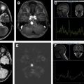
As a marker of neuronal density, NAA level decreases in all types of gliomas with an inverse correlation between NAA level and tumor grade. Decrease in NAA level is associated with infiltration and destruction of neurons by tumoral tissue. Investigated by numerous studies, increased Cho (a marker of cell membrane turnover) concentration occurs in all grades of gliomas. The relative signal intensity of Cho and Cho/Cr ratio increases significantly from low-grade toward high-grade gliomas ( Figs. 3 and 4 ). A strong correlation is described between rCBV with ADC and choline level in glioblastomas. Increased choline concentration (>45%) on serial MRS is described as an indicator of malignant transformation in low-grade gliomas. However, characterization of gliomas based on choline index should be made with caution. Shimizu and colleagues reported a powerful correlation of choline level and Ki-67 index in gliomas with homogeneous MR imaging pattern, but not in heterogeneous gliomas. They suggested that choline is not reliable for predicting malignancy in heterogeneous gliomas. In glioblastomas with extensive necrosis and less viable cells, Cho/Cr and Cho/NAA ratios are diminished, but areas with high cellular density show Cho/Cr ratios greater than 2 in these high-grade tumors. In contrast, absence of choline peak does not exclude CNS glioma.


