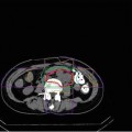
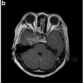
Fig. 32.1
(a) Axial computed tomography before carbon ion radiotherapy. (b) Representative axial isodose distributions. 11C-labeled methionine positron emission tomography images (c) before carbon ion radiotherapy and (d) 40 months after treatment
32.5.2 Case 2 (Sacral Chordoma)
A 33-year-old man with sacral chordoma underwent surgical resection followed by postoperative radiotherapy administered at 50 Gy in 25 fractions. Twenty years later, he developed local recurrence, which was confirmed by biopsy. He was referred to our hospital for C-ion RT. The tumor located at the edge of the residual sacrum had a maximum diameter of 60 mm (Fig. 32.2a, b). He was immobilized in the prone position and irradiated using three portals. C-ion RT was delivered at 64.0 GyE in16 fractions. The critical organs such as the colon and skin were spared (Fig. 32.2c, d). In addition, the bilateral sciatic nerves could be spared. Seven years later, the patient was disease-free and could walk without an aid.
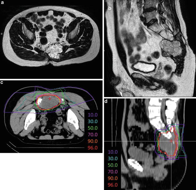

Fig. 32.2
(a) Axial and (b) sagittal T2-weighted magnetic resonance images obtained before carbon ion radiotherapy. Representative (c) axial and (d) sagittal isodose distributions
32.5.3 Case 3 (Myxoid Liposarcoma in the Left Buttock)
Stay updated, free articles. Join our Telegram channel

Full access? Get Clinical Tree


