div style=”display:none;”> VISUAL CORTEX: LOCATION AND COACTIVATION VISUAL CORTEX: FUNCTIONAL CONNECTIVITY FUNCTIONAL CONNECTIVITY TO VISUAL CORTEX VISUAL CORTEX: LOCATION AND SUBREGIONS
Primary Visual and Visual Association Cortex (Areas 17, 18, 19)
Main Text
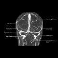
Location and Boundaries
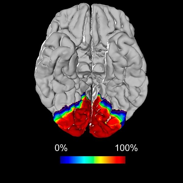
Location
Boundaries
Function
Vision
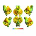
Structural Connections
Cortical
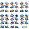
Functional Connections
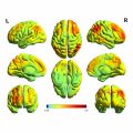
Coactive Regions
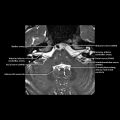
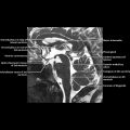
Image Gallery
Print Images


Additional Images


![]()
Stay updated, free articles. Join our Telegram channel

Full access? Get Clinical Tree


Primary Visual and Visual Association Cortex (Areas 17, 18, 19)