Principles of Venous Access


Principles of Venous Access


David O. Kessel and Karen Flood


Clinical Relevance


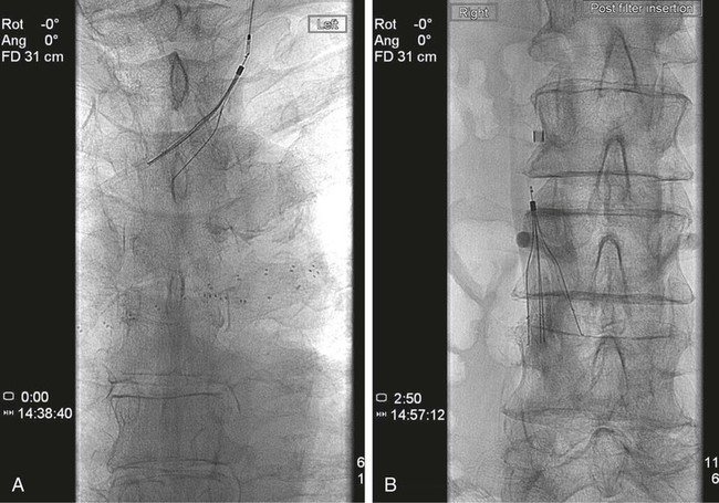
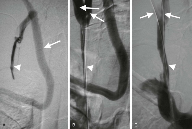
The National Institute of Clinical Excellence has recommended that ultrasound guidance should be used when inserting elective central venous lines.1 There is clear evidence that using imaging guidance increases success rates and reduces complications associated with jugular and subclavian vein puncture. Most of these routine procedures do not require interventional radiologic input, but interventional radiologists become indispensable in cases where central venous access is difficult, as is often the case in hemodialysis and cancer patients.2


Indications


Venous access is the starting point for a large number of diagnostic and therapeutic interventions in the systemic and portal venous circulations. Table 29-1 lists typical indications.


Contraindications


There are few absolute contraindications to obtaining venous access. Reversal of coagulopathy should be considered if clotting is severely deranged. This is particularly important if solid organ puncture is required or large-bore catheters are being used.


Venous thrombosis, stenosis, and occlusion are frequently encountered but are not contraindications to access. Rather, these are the reasons an interventional radiologist (i.e., an individual with skill in thrombolysis and recanalization of vessels, etc.) is required. With regard to venous thrombosis and risk of causing emboli, care should be taken to avoid puncturing into iliofemoral deep venous thromboses (DVT) when placing an inferior vena cava (IVC) filter. This is one reason for considering the jugular route as a standard approach for IVC filter placement.


Equipment


Ultrasound


The key item of equipment is an ultrasound machine with a suitable-frequency probe (typically 4-9 MHz for jugular vein puncture and 3-5 MHz for transhepatic, transrenal, or transsplenic puncture) and color Doppler. Poor-quality ultrasound is the most common cause of failure to obtain venous access. Certain compact ultrasound systems such as those used in wards for venous puncture are often of insufficient quality for this purpose.


Angioplasty Balloons and Stents


It is sometimes necessary to predilate a stenosed or occluded vein prior to passage of a line (Fig. 29-1). Stents are generally unnecessary, however, if access rather than patency is the desired outcome.


image
FIGURE 29-1 Insertion of Tesio lines (Medcomp, Harleysville, Pa.) through an occluded jugular vein (same patient as Fig. 29-3). A, Remnant of jugular vein (arrowhead) and collateral vein passing into subclavian vein (arrow). B, Occluded internal jugular vein has been recanalized and a guidewire passed from each of two veins (arrows) cranial to occlusion. C, Following balloon angioplasty, jugular vein is now patent, and collateral vein seen in A no longer fills. Lines were inserted uneventfully.


Technique


Veins are typically thin walled and perfused at low pressure. These characteristics increase the challenge of puncture. To improve the odds, it helps to increase the venous pressure. A tourniquet is fine in the periphery but is of little use in the jugular vein. The Valsalva maneuver will distend a vein if the patient is able to follow the instruction to “strain gently down.” It is often helpful to ask the patient to hum to achieve this desired effect. Ensuring the patient is adequately hydrated can also help, and if necessary, administering a liter of intravenous saline will also work to increase the venous pressure. An alternative is to use the head-down (Trendelenburg) position or temporarily elevate the patient’s legs to increase venous return. Unfortunately, few interventional radiology suites are equipped with tilting tables, so Trendelenburg is often not an option. Interventionists will also typically be referred all patients who cannot lie flat for one reason or another, so it is important that he or she be familiar with the different means of increasing venous pressure in these patients.


Whatever technique is adopted, one should be sure that the final positioning allows scanning the vein while performing the puncture. It is best to choose an approach that allows the needle to pass along the scan plane so it can be seen throughout its passage, but this is often impossible when using a standard linear array probe to guide jugular puncture because there is insufficient space in the neck. In these circumstances, it is necessary to scan transversely and “triangulate” the needle position as it approaches the vein. It is worth remembering that thin-walled veins will often be compressed by pressure transmission from the needle in soft tissues superficial to or even adjacent to the vein. This is no substitute for observing the needle tip come into contact with the anterior vein wall. When the needle actually abuts the vein, the wall will be indented by the needle, and the anterior wall may even come into contact with the posterior wall. Further needle advancement in this circumstance leads to transfixing the vein. To puncture the anterior wall and enter the lumen, it is best to advance the needle with a short stab as soon as it abuts the anterior wall. If a syringe was attached at the start of the procedure, blood should be aspirated to confirm intraluminal position. If aspiration is not possible, it is likely the needle has been advanced through the posterior vein wall. In this case, the needle should be pulled back slowly, aspirating as it goes, until flow is restored. If this does not succeed, one should start again, ensuring the needle is flushed before beginning.


Anatomy and Approach


Several considerations may be relevant when planning the optimal approach for venous access. The general adage that the shortest, straightest route is best often applies, but this should be weighed against possible disadvantages of choosing a particular site. Typical approaches are outlined in Table 29-1, but there are also some esoteric routes typically reserved for cases wherein all other options have failed. These include utilizing collateral veins, transhepatic, translumbar, or transrenal access to the systemic veins and transsplenic or transmesenteric access to the portal venous system. There are occasions when dual access is helpful, especially when the most direct route is through a small vessel or via an artery.


Virtually any vein can be used, but the most common sites depend on the indication. The following section will outline a rational approach for the most common procedures.


Central Venous Access


Central venous access is the starting point for many procedures (see Table 29-1). The most common points of access are the internal jugular and subclavian veins. The jugular vein is superior to the subclavian vein because it is less prone to symptomatic thrombosis.3 The right internal jugular vein also provides the shortest, straightest route and is the first-choice point of access. The left jugular approach is often possible, but it is important to be aware of any sharp bends in the mediastinum that may result in kinking of sheaths (Fig. 29-2). It is prudent to check the manufacturer’s instructions for use; there are sometimes caveats to using the left jugular approach for this reason.


Stay updated, free articles. Join our Telegram channel

Dec 23, 2015 | Posted by in INTERVENTIONAL RADIOLOGY | Comments Off on Principles of Venous Access

Full access? Get Clinical Tree

Get Clinical Tree app for offline access