Fig. 1
Round small solid pseudopapillary tumor. (a) Surgical specimen (intermediate pancreatectomy). (b–d) Ultrasound study: B-mode US shows a small solid hypoechoic lesion in the pancreatic tail, with well-defined margins (b). Color Doppler US does not show any intralesional vessels (c). At CEUS, the lesion shows rim enhancement (d). (e, f) MRI study: the lesion appears slightly hyperintense on T2-weighted MRI images (e), and enhancement of the tumor capsule is seen on T1-weighted images (f)
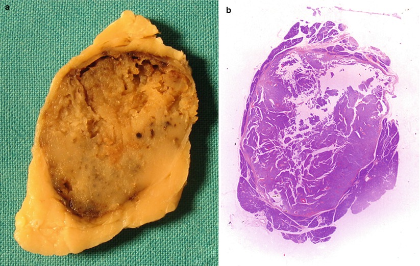
Fig. 2
Solid pseudopapillary tumor. (a, b) Surgical specimen: the well-demarcated neoplasm appears mostly solid on the cut section (a). The whole mount macrosection shows that the lesion is truly not solid, but there are initial cystic degenerative changes with small pseudopapillae (b)
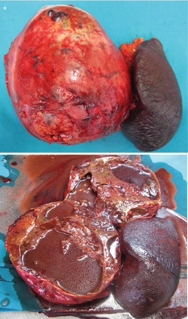
Fig. 3
Large solid pseudopapillary tumor with extensive hemorrhage and necrotic changes. (a, b) Surgical specimen (distal pancreatectomy): large round lesion in the body-tail of the pancreas (a). This is an example of almost entirely cystic tumor: the cut specimen shows extensive hemorrhage and necrotic changes (b)
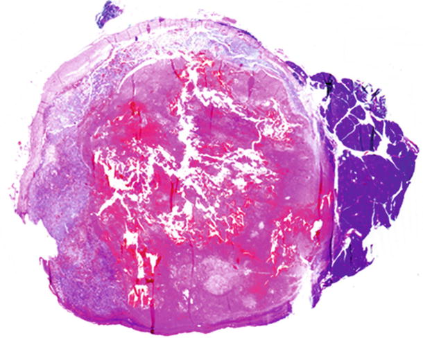
Fig. 4
Solid pseudopapillary tumor with extensive hemorrhagic changes. Whole mount macrosection shows extensive hemorrhagic changes
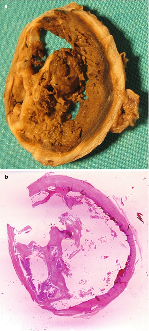
Fig. 5
Solid pseudopapillary tumor with large hematoma. (a, b) Surgical specimen: a large hematoma fills the neoplastic cavity (a) of this cystic lesion (b)
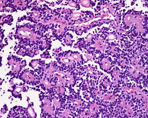
Fig. 6
Solid pseudopapillary tumor. Histopathology: prominent pseudopapillary pattern of the tumor: a variable number of loosely cohesive cells surround delicate blood vessels (Courtesy of Piccin Editore, Milan, Italy)
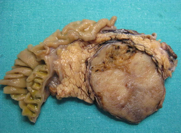
Fig. 7
Large solid pseudopapillary tumor. Surgical specimen (pancreaticoduodenectomy): large tumor of the pancreatic head, with expansive growth and mainly solid appearance
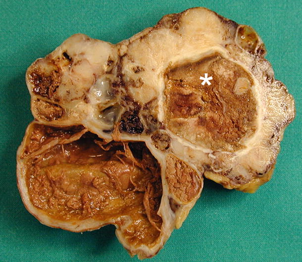
Fig. 8
Solid pseudopapillary tumor. Surgical specimen (distal pancreatectomy): solid and cystic components with areas of recent and remote hemorrhage (asterisk)
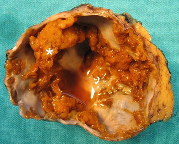
Fig. 9
Large cystic pseudopapillary tumor. Surgical specimen (distal pancreatectomy): large predominantly cystic lesion; some grossly visible vegetations can be seen (asterisk)
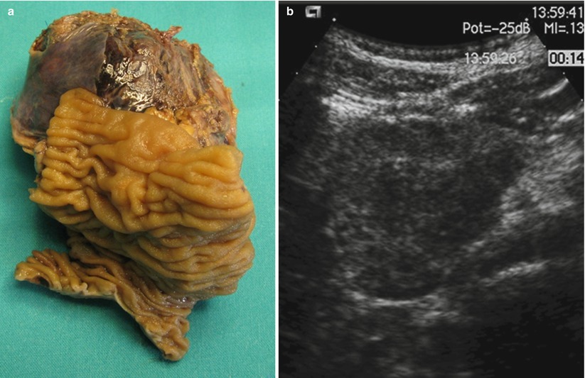
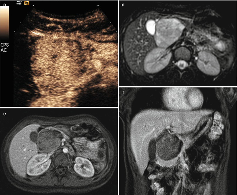
Fig. 10
Solid pseudopapillary tumor. (a) Surgical specimen (pancreaticoduodenectomy): large round well-defined tumor of the pancreatic head. (b, c) ultrasound study: it appears homogeneously hypoechoic at baseline US (b); at CEUS, a small cystic central portion is depicted (c). (d–f) MRI study: the lesion appears inhomogeneously hyperintense on T2-weighted MRI images (d) and shows poor enhancement after Gd-administration (e, f)
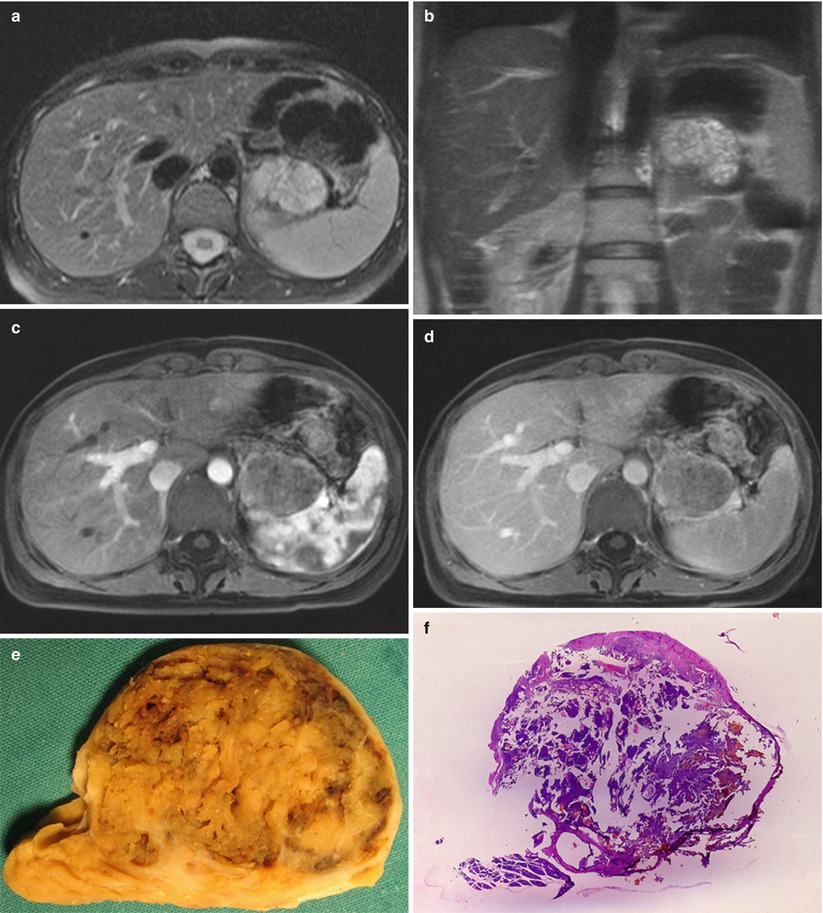
Fig. 11
Solid pseudopapillary tumor. (a–d) MRI study: axial (a) and coronal (b) T2-weighted MRI images show a large well-demarcated lesion, which appears inhomogeneously hyperintense. The lesion shows peripheral enhancement after Gd-administration, both in the arterial (c) and venous (d) phases. (e, f) Surgical specimen (distal pancreatectomy): a large well-demarcated lesion with gross pseudopapillae (e), which are also present in the whole mount macrosection (f)
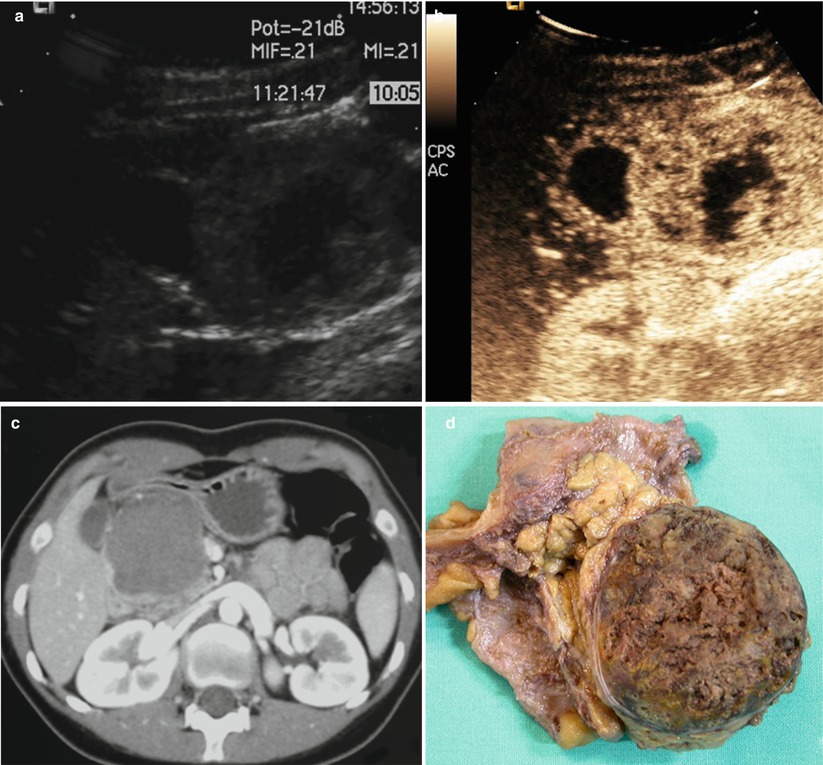
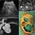
Fig. 12
Pseudopapillary tumor with large cystic area. (a, b) Ultrasound study: B-mode US shows a large round well-demarcated lesion in the pancreatic head, which appears hypoechoic with a large central fluid area (a). CEUS (b) confirms the presence of solid papillae and fluid areas. (c) CT study: axial CECT shows a large hypodense lesion with lobulated margins and peripheral enhancement. (d) Surgical specimen (pancreaticoduodenectomy): the cut section reveals soft, friable brownish neoplastic tissue
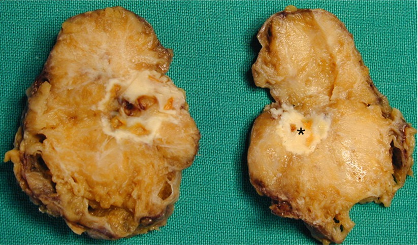
Fig. 13
Pseudopapillary tumor. Surgical specimen (distal pancreatectomy): large, mostly solid, tumor with paracentral gross calcification (asterisk) and small cystic areas in the periphery of the tumor
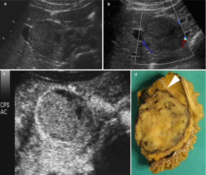
Fig. 14
Solid round pseudopapillary tumor. (a–c) Ultrasound study: B-mode US (a) shows a large round well-demarcated lesion in the pancreatic head (calipers in a), predominantly solid, with some cystic portions. Color Doppler US (b) shows no intratumoral signal. The lesion appears well-vascularized at CEUS (c). (d) Surgical specimen (pancreaticoduodenectomy): solid round well-demarcated tumor of the pancreatic head. The tumor displaces, but does not infiltrate, the main pancreatic duct (arrowhead in d)
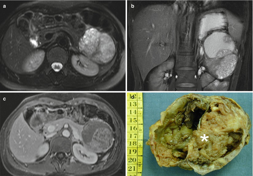
Fig. 15
Large pseudopapillary tumor. (a–c) MRI study: axial (a) and coronal (b) T2-weighted images show a large lesion in the pancreatic tail, inhomogeneously hyperintense, with cystic portions of different sizes. Axial T-1 post-Gd image shows enhancing portions (c). (d) Surgical specimen (distal pancreatectomy): the tumor shows prominent cystic degeneration, with large pseudopapillae (asterisk) (Courtesy of Piccin Editore, Milan, Italy)
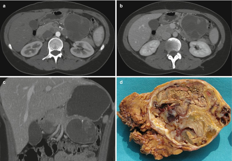
Fig. 16
Large pseudopapillary tumor. (a–c) CT study: large well-demarcated tumor in the pancreatic tail, with rim enhancement in the arterial (a) and venous (axial b, coronal c) images. (d) Surgical specimen (distal pancreatectomy): the cut section shows yellow-brownish solid areas and irregularly shaped cystic components with areas of recent and remote hemorrhage
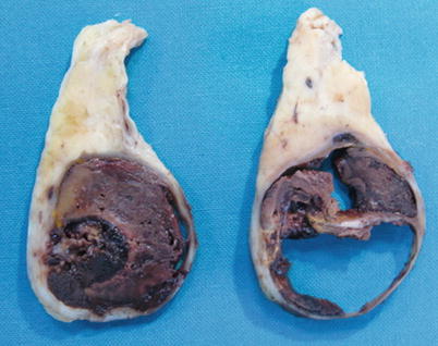
Fig. 17


Mucinous cystic neoplasm resembling a solid pseudopapillary tumor. Surgical specimen: the cystic lesion is completely occupied by a large hematoma
Related posts:


Stay updated, free articles. Join our Telegram channel
Full access? Get Clinical Tree
