Fig. 66.1
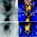
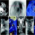
PET-CT images with parenchymal window show a left solid, uneven, apical lung nodule, with branches infiltrating the surrounding parenchyma (yellow arrow). There are also multiple lymph nodes of increased size (blue arrow) and a metastasis at the level of a dorsal vertebra (red arrow). All of these lesions have high metabolism
66.4 Conclusions
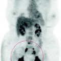
The PET scan suggests left lung cancer with multiple lymph node and bone metastases.
Histological definition of pulmonary nodule is suggested.
Less likely is the possibility of metastasis of breast cancer operated in 2001. See Figs. 66.2, 66.3.
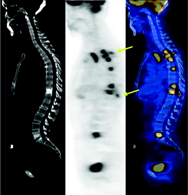

Fig. 66.2


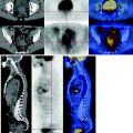
The sagittal CT scan reconstruction of the spine shows thickening of two vertebral bodies, D11 and L5 that at the PET scan have high metabolism. There were no thickening morphostructural alterations at D3, D4, D5, D7, D10 and some spinous processes (D3, D11), which, however, are characterized by high consumption of glucose. This finding suggests the presence of bone marrow metastases that have not yet determined a bone producing skeletal alteration in, an event that is realized only belatedly
Stay updated, free articles. Join our Telegram channel

Full access? Get Clinical Tree


