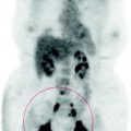
Fig. 60.1
MIP image: The high lung deposition of FDG is determined by the condition of chronic inflammation of the interstitium and by the presence of lymphocytic alveolitis
The extent of the interstitial disease in this patient seems remarkable: the pulmonary uptake exceeds that of the mediastinum and liver.
As the gallium scintigraphy, PET-FDG allows us to evaluate the biological activity of the disease, while the CT shows the morphology of the fibrotic damage.
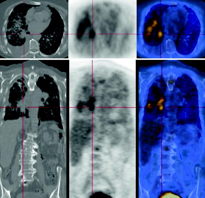
Fig. 60.2
 Laryngeal Squamous Carcinoma: Staging
 Radio-Treated Cancer of the Posterior Hemi-Circumference of the Anal Canal: Post-Actinic Fibrosis
 Bone-Destroying Metastases in Thyroid Undifferentiated Carcinoma
 Surgically Treated Endometrial Cancer: Bilateral Nodal Recurrence
 Undifferentiated Gastric Adenocarcinoma with Peritoneal Carcinosis
 Bone Metastases from Breast Cancer: Progression of Disease and Subsequent Response to Radiotherapy


At CT, a marked reticular thickening of the interstitium, diffusely involving the parenchyma of both lungs, can be seen. At the inferior bronchus of the right lung, a ground-glass nodule is seen, characterized by ill-defined margins and satellite areas of parenchymal consolidation. The FDG-PET demonstrates limited consumption of glucose in the lesion
Related posts:
Laryngeal Squamous Carcinoma: Staging
Radio-Treated Cancer of the Posterior Hemi-Circumference of the Anal Canal: Post-Actinic Fibrosis
Bone-Destroying Metastases in Thyroid Undifferentiated Carcinoma
Surgically Treated Endometrial Cancer: Bilateral Nodal Recurrence
Undifferentiated Gastric Adenocarcinoma with Peritoneal Carcinosis
Bone Metastases from Breast Cancer: Progression of Disease and Subsequent Response to Radiotherapy
Stay updated, free articles. Join our Telegram channel
Full access? Get Clinical Tree
