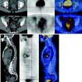
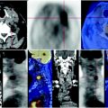
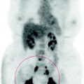
Fig. 69.1
At the posterior segment of the upper lobe of the left lung, CT-PET shows a parenchymal nodule with ill-defined margins, at the basis of the parietal pleura, that seems to infiltrate adjacent tissues due the presence of spikes. The biological activity of the lesion is low
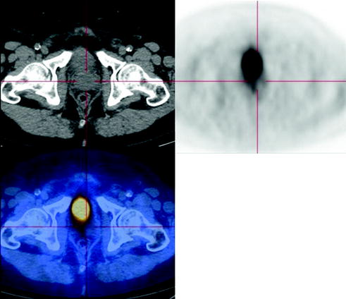
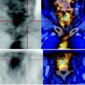
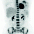
Fig. 69.2
The PET-CT scan shows a slightly increased prostate in size, with inhomogeneous density. There were no focal lesions characterized by high metabolism because prostatic adenocarcinomas have little affinity for FDG
69.5 Key Points
In patients without a known malignancy, the presence of multiple pulmonary nodules ≥ 1 cm in diameter is most commonly due to metastatic lesions, and nodules < 5 mm in diameter, which overlap the visceral pleura or interlobular septa and are detected incidentally, are most commonly benign lesions.
Related posts:
 Laryngeal Squamous Carcinoma: Staging
 Radio-Treated Cancer of the Posterior Hemi-Circumference of the Anal Canal: Post-Actinic Fibrosis
 Bone-Destroying Metastases in Thyroid Undifferentiated Carcinoma
 Surgically Treated Endometrial Cancer: Bilateral Nodal Recurrence
 Metastatic Breast Carcinoma: Restaging After Neoadjuvant Chemotherapy
 Bone Metastases from Breast Cancer: Progression of Disease and Subsequent Response to Radiotherapy
Stay updated, free articles. Join our Telegram channel

Full access? Get Clinical Tree


