- •
Size of the pulmonary annulus in comparison with the aortic annulus.
- •
Laminar or turbulent flow across pulmonic valve?
- •
Peak systolic velocity across the pulmonic valve.
- •
Morphology of the pulmonary valve.
- •
Size of the tricuspid annulus compared with the mitral annulus.
- •
Degree of tricuspid regurgitation.
- •
Right ventricle pressure estimate based on tricuspid regurgitation peak velocity.
- •
Right atrial size.
- •
Right ventricle size and function.
- •
Degree of right ventricular hypertrophy.
- •
Direction of flow within the ductus arteriosus.
- •
Size of the branch pulmonary arteries.
- •
Size and appearance of the foramen ovale.
- •
Evidence for elevated central venous pressure as marked by reversal of flow with atrial contraction in the ductus venosus.
- •
Evidence for elevated central venous pressure as marked by pulsations within the inferior vena cava.
- •
Presence of any ventriculocoronary “sinusoid” connections.
Anatomy and Anatomical Associations
Pulmonary stenosis is a heterogeneous disorder. The obstruction may occur at the subvalvar, valvar, or supravalvar levels ( Figure 24-1 ). In subvalvar stenosis, the obstruction may be caused by either muscle or fibrous tissue. Muscular subvalvar stenosis includes obstruction secondary to malalignment of the conal septum or hypertrophic cardiomyopathy. Fibrous subvalvar stenosis typically occurs in association with conotruncal abnormalities and may be caused by abnormal chordal attachments or abnormal atrioventricular valve leaflet attachments.

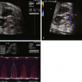
Valvar pulmonary stenosis may be classified into six anatomic subtypes: (1) doming (42%), (2) unicommissural (16%), (3) bicuspid (10%), (4) tricuspid (6%), (5) hypoplastic annulus (6%), and (6) dysplastic (19%). In doming pulmonary stenosis, the valve appears dome-shaped or conical with a small central orifice varying in size from a pinhole to several millimeters in diameter. There is no separation into leaflets in doming pulmonary stenosis. In unicommissural pulmonary valve stenosis, there is a single commissure (natural split between the leaflets) and a single thickened leaflet. In bicuspid pulmonary valve stenosis, two commissures and two thickened leaflets are present. In tricuspid pulmonary valve stenosis, there is moderate thickening of all three leaflets and partial fusion of all three commissures. In hypoplastic pulmonary valve stenosis, the annulus is markedly narrowed. Finally, in dysplastic pulmonary valve stenosis, the three leaflets are markedly thickened and redundant, although the commissures are not fused. Dysplastic pulmonary valves are present in the majority of cases of Noonan’s syndrome and in some nonfamilial cases of pulmonary stenosis. In supravalvar pulmonary stenosis, the obstruction occurs above the annulus of the pulmonary valve.
Pulmonary stenosis may be seen in association with a number of different types of congenital heart disease, including conotruncal abnormalities such as tetralogy of Fallot, double-outlet right ventricle or transposition of the great arteries, ventricular septal defect, tricuspid atresia or stenosis, Ebstein’s anomaly, and congenitally corrected transposition of the great arteries. With more severe degrees of valvar obstruction, the infundibulum (conus) of the right ventricle may become hypertrophied, producing dynamic subvalvar stenosis. Poststenotic dilation of the main pulmonary artery may occur on account of the high-velocity jet of flow being ejected through the stenotic pulmonary orifice. Tricuspid regurgitation may occur secondary to increased right ventricular pressure. The right atrium may dilate secondary to elevated filling pressures within the hypertrophied right ventricle and altered compliance. Smallness or hypoplasia of the right ventricle can be seen in association with isolated, but severe, pulmonic stenosis. Finally, in severe degrees of obstruction seen in the fetus, in the absence of significant tricuspid regurgitation, connections between the hypertensive right ventricle cavity and the developing coronary circulation—coronary sinusoids—may form. Abnormalities within the coronary circulation, including stenoses, interruptions, or even atresia of the origin of the coronary artery, place the fetus at risk for myocardial ischemia and infarction within the subendocardium of the right ventricle. Although these can be seen in severe pulmonic stenosis, they are more common in the presence of pulmonary atresia.
Frequency, Genetics, and Development
Isolated pulmonary valve stenosis occurs in 7% to 12% of patients with congenital heart disease. In contrast, if associated lesions are included, pulmonary stenosis occurs in 25% to 30% of all patients with congenital heart disease. Familial studies suggest a 2.1% incidence of congenital heart disease, usually either tetralogy of Fallot or pulmonary stenosis, in siblings of patients with pulmonary stenosis. If a parent has pulmonary stenosis, the incidence of pulmonary valve stenosis in a child is 2.8%. In twin studies, 8.3% of monozygotic twins and 2.2% of dizygotic twins had pulmonary stenosis.
Genetic syndromes may be associated with pulmonary stenosis. These include neurofibromatosis, multiple lentigines or LEOPARD (lentigines, electrocardiogram abnormalities, ocular hypertelorism, pulmonary stenosis, abnormal genitalia, retardation of growth, and deafness) syndrome, and Noonan’s syndrome. Up to 50% of patients with Noonan’s syndrome have a congenital heart defect, usually a dysplastic pulmonary valve, and up to 25% of patients with Noonan’s syndrome have hypertrophic cardiomyopathy, which may also be associated with pulmonary stenosis. In Williams’s syndrome, supravalvar pulmonary stenosis can be seen. Systemic diseases such as glycogen storage disease may be associated with infundibular and valvar pulmonary stenosis.
Prenatal Physiology
Fetal physiology depends upon the severity of the pulmonary valve stenosis. Mild forms of pulmonary stenosis are well tolerated in utero and may not even be detected by screening fetal echocardiography. Diagnosis depends upon careful inspection of pulmonary valve morphology and pulse wave Doppler interrogation of the pulmonary valve. A peak velocity greater than 1 m/sec across the pulmonary valve is abnormal and should alert the clinician to the possibility of pulmonary valve stenosis. With moderate to severe pulmonary stenosis, right ventricular hypertrophy, tricuspid regurgitation, and poststenotic dilation of the main pulmonary artery may be seen. Flow within the ductus arteriosus may be reversed (aorta–to–pulmonary artery) in order to augment pulmonary blood flow. With severe degrees of obstruction, right ventricular filling pressures may be high, leading to increased right-to-left shunting across the patent foramen ovale and dilation of the left side of the heart as a consequence. Right ventricular hypoplasia to varying degrees can also be seen in such cases. As blood is diverted away from the right ventricle, growth is impaired. Normally, the tricuspid valve annulus is larger than the mitral valve annulus throughout gestation. In severe pulmonic stenosis, the right ventricular cavity and tricuspid valve annulus may not grow at an expected rate, leading to the finding of a mitral valve annulus that is larger than the tricuspid.
In “critical” pulmonary stenosis in the absence of significant tricuspid regurgitation, the right ventricular pressure may become even higher than that in the left ventricle (suprasystemic pressure). Coronary sinusoids may form from the right ventricular cavity to the coronary circulation. Abnormalities within the coronary circulation, including stenoses, interruptions, or even atresia of the origin of the coronary artery, places the fetus at risk for myocardial ischemia and infarction.
Prenatal Management
Family counseling depends upon the severity of the obstruction. Mild to moderate cases of pulmonary stenosis are well tolerated in utero and typically do not require neonatal intervention. However, pulmonary valve stenosis may progress over the course of gestation from a milder to a more severe form. Consequently, serial surveillance over the course of gestation is appropriate. Severe cases of pulmonary stenosis will require neonatal intervention, either in the cardiac catheterization laboratory or in the operating room.
A number of important features are to be imaged and assessed at initial diagnosis and at subsequent prenatal visits when pulmonary stenosis is suspected (see Key Features). Vaginal delivery is generally well tolerated by most fetuses with pulmonary stenosis. However, cesarean section may be recommended for fetuses with critical pulmonary stenosis if impending hydrops fetalis is suspected, if there is evidence for left ventricular dysfunction, or if there is a restrictive patent foramen ovale. Prostaglandin should be recommended at birth if there is inadequate antegrade flow across the pulmonary valve and reversal of flow within the ductus arteriosus.
Most fetuses with pulmonary stenosis have perhaps a somewhat small but adequate size right ventricle and achieve a biventricular repair after birth. Consequently, prenatal intervention is usually not recommended. However, some centers have advocated balloon valvuloplasty in utero in cases of critical pulmonary stenosis in order to prevent the development of hydrops fetalis and to ensure a biventricular circulation postnatally. Indications cited for in utero intervention include impending hydrops fetalis, marked by the appearance of ascites, pericardial, or pleural effusions, decreased left ventricular shortening, marked flow reversal with atrial contraction within the ductus venosus, pulsations within the inferior vena cava, and restrictive flow across the patent foramen ovale.
Related posts:


Stay updated, free articles. Join our Telegram channel

Full access? Get Clinical Tree


