Pyogenic Discitis and Epidural Abscess
Benjamin Y. Huang
CLINICAL HISTORY
42-year-old patient with a history of IV drug abuse presenting with low back pain, which has progressively worsened over several weeks.
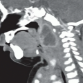
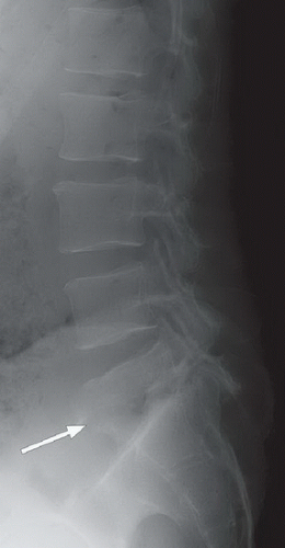 FIGURE 81A |
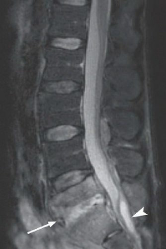 FIGURE 81B |
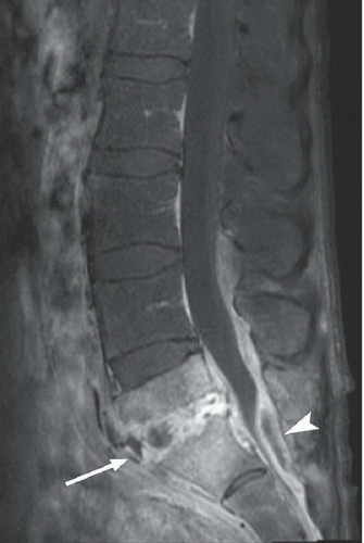 FIGURE 81C |
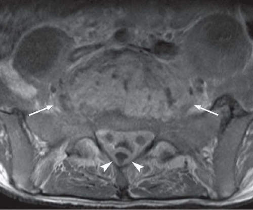 FIGURE 81D |
FINDINGS:
Figure 81A: Lateral radiograph of the lumbar spine demonstrates severe collapse of the L5-S1 intervertebral disk space (arrow) with erosion of the inferior L5 endplate and superior S1 endplate. Figure 81B: Sagittal STIR image demonstrates abnormal hyperintense signal within the disk space (arrow) and throughout the adjacent L5 and S1 vertebral bodies. There is also a lentiform fluid collection posterior to the thecal sac (arrowhead), which appears to displace the dura anteriorly. Figures 81C and 81D: Corresponding contrast-enhanced sagittal, fat-suppressed T1-weighted image (Fig. 81C), and contrast-enhanced axial T1-weighted image at L5-S1 (Fig. 81D) demonstrate
enhancement of the L5-S1 disk space (arrows) and the adjacent vertebral bodies. In addition, there is abnormal enhancement in the epidural and prevertebral soft tissues, including portions of the psoas muscles. Also note enhancement along the periphery of the aforementioned epidural fluid collection (arrowheads) which is compressing the spinal canal.
enhancement of the L5-S1 disk space (arrows) and the adjacent vertebral bodies. In addition, there is abnormal enhancement in the epidural and prevertebral soft tissues, including portions of the psoas muscles. Also note enhancement along the periphery of the aforementioned epidural fluid collection (arrowheads) which is compressing the spinal canal.
Related posts:


Stay updated, free articles. Join our Telegram channel

Full access? Get Clinical Tree


