Spinal infections are a spectrum of disease comprising spondylitis, diskitis, spondylodiskitis, pyogenic facet arthropathy, epidural infections, meningitis, polyradiculopathy, and myelitis. Inflammation can be caused by pyogenic, granulomatous, autoimmune, idiopathic, and iatrogenic conditions. In an era of immune suppression, tuberculosis, and HIV epidemic, together with worldwide socioeconomic fluctuations, spinal infections are increasing. Despite advanced diagnostic technology, diagnosis of this entity and differentiation from degenerative disease, noninfective inflammatory lesions, and spinal neoplasms are difficult. Radiological evaluations play an important role, with contrast-enhanced MR imaging the modality of choice in diagnosis, evaluation, treatment planning, interventional treatment, and treatment monitoring of spinal infections.
Key points
- •
With increasing immune suppression and globalization, spinal pyogenic infections are increasing in incidence.
- •
MR imaging is the modality of choice in the imaging work-up of pyogenic spinal infections.
- •
Characteristic imaging findings together with microbiological isolation of the causative agent are the main tools for appropriate diagnosis.
- •
Radiologists play an important role, because early diagnosis and treatment monitoring are extremely important in preventing morbidity and fatality.
Introduction
Pyogenic spinal infection is a life-threatening neurologic condition encompassing a broad range of clinical entities, including spondylitis, spondylodiskitis, septic diskitis, pyogenic facet arthropathy, epidural infection-abscess, leptomeningitis, and myelitis-spinal cord abscess. As a significant cause of morbidity and mortality, it is difficult to differentiate from degenerative processes, inflammatory disorders, metabolic disorders, and neoplasms. Pyogenic spinal infections have a reported incidence of 0.2 to 2 cases per 100.000 per year. Men seem to be affected twice as often as women, with a peak incidence in the sixth decade. Globalization, improved life expectancy, comorbid factors, such as diabetes mellitus, drug abuse, overuse of antibiotics, immune suppression, malignancy, chronic diseases, spinal instrumentation, and increased awareness together with highly performing advanced imaging techniques have led to a rising incidence and frequency of diagnosis of spinal infections. Hematogenous spread, direct inoculation, and contiguous spread are the main routes of spinal infection. The infection is generally unimicrobial, with Staphylococcus aureus the main causative agent. Characteristic imaging findings together with microbiological isolation of the causative agent are the main tools for appropriate diagnosis. Radiographs, CT, scintigraphy, positron emission tomography (PET)-CT, and MR imaging can all be used for this purpose and for confirmation and localization.
Spondylitis and spondylodiskitis
Background and Pathophysiology
Pyogenic spondylitis/spondylodiskitis is a bacterial infection of the spinal column, intervertebral disks in association with paraspinal soft tissue, epidural space, and/or ligaments of the extradural spine infection-extension. The infection is usually unimicrobial. S. aureus accounts for approximately 60% and Enterobacter for 30% of cases. Klebsiella , Pseudomonas , Serratia , and Salmonella (in patients with sickle cell anemia) are other common organisms. S. aureus is known to produce hyaluronidase, which is a proteolytic enzyme causing lysis of the disk. The infection source is generally from a urinary tract, pulmonary, pelvic, but also can be from a cutaneous infection, intravenous (IV) injections with contaminated needles, cellulitis, fasciitis, subcutaneous abscess, or pyomyositis. Predisposing factors include diabetes mellitus; other chronic diseases, such as renal failure and cirrhosis; immunosuppressed states; and IV drug use. Organisms reach the spine by 2 main routes:
- 1.
Hematogenous spread
- 2.
Nonhematogenous spread
Hematogenous spread can be arterial or venous. Arterial spread is more common, as a result of septicemia, and occurs at the end arteriolar level supplying the vertebral body. In adults, the disk is avascular. Hematogenous organisms arrive in vertebrae via end arteriolar arcades of metaphyseal equivalent areas. Those areas correspond to subchondral plate adjacent to the disk, particularly in the anterior part. Through the disruption of cortical bone, organisms can extend to subligamentous paravertebral epidural spaces, disk, and contiguous vertebrae. Secondary infection, however, may occur in degenerative disk disease. Granulation tissue, with the ingrowth of vessels, may penetrate radial tears and make direct hematogenous spread of infection of the disk in these cases. In children, persisting vascular channels may allow direct inoculation of the disk; thus, children initially may present with diskitis alone. Because of stretching of the anterior longitudinal ligament from diskitis, abdominal pain can be the initial presenting symptom in children.
The transvenous route is another hematogenous spreading path. The epidural venous plexus within the central canal, the Batson plexus, represents a series of valveless veins that extends the length of the spinal canal. The venous spreading to the spine is of particular importance in the urinary tract and other pelvic organ infections. Because the Batson plexus is a valveless system, increasing intra-abdominal pressure allows retrograde hematogeneous spread from the pelvis and abdominal organs to the vertebral column.
Nonhematogeneous spread consists of penetrating trauma, direct exposure related to skin breakdown or open wounds, surgical procedures, and diagnostic interventions, such as lumbar puncture, epidural block, nerve block, vertebroplasty, or catheterization. Compared with hematogeneous spread, direct inoculation has a prominent predilection for the pedicle, laminae, and spinous processes.
Clinical Presentation
The diagnosis of pyogenic spondylodiskitis relies on clinical, imaging, and laboratory findings. Spinal involvement level varies, and infections have been reported at all spinal segments. The anatomic site of predilection is the lumbar region, with a frequency decreasing caudocranially over the spinal column. Usually there is a delay of 2 to 12 weeks in diagnosis, which can lead to bony destruction, kyphosis, and subsequent neurologic complications. Depending on the location and extent of the infectious process, symptoms and neurologic deficits may vary. Persistent back pain aggravated by motion, malaise, fever, anorexia, tenderness, and rigidity may be the presenting symptom. A more insidious onset with only back pain and discomfort is also possible. On physical examination, signs of nerve root compression with radiculopathy, meningeal irritation, lower extremity weakness, or paraplegia can be present with epidural involvement. Difficulty in swallowing is also another symptom in patients with cervical pyogenic spondylitis and retropharyngeal abscess. If untreated, there is an overall mortality ranging between 18% and 31%.
Laboratory Tests
An initial work-up consisting of white blood cell (WBC) count, erythrocyte sedimentation rate (ESR), C-reactive protein (CRP) level, Gram stain, and blood culture should be performed for each patient suspected of pyogenic spondylitis. Together with blood culture and Gram staining, elevated ESR and CRP levels are reported as good markers of infection. ESR is found elevated in 70% to 100% of infections, and average ESR in patients with pyogenic spondylitis ranges between 43 and 87 mm per hour. Elevated ESR, however, is not specific for infection and its use in following disease progression should be cautioned because it normalizes in an irregular and slow fashion, even after successful treatment. WBC count is elevated in only 13% to 60% of cases in a moderate fashion. Although not crucial in the diagnosis, WBC count can provide general guidance in evaluating treatment response. Knowing that pyogenic spondylodiskitis has a variable source of infection, blood culture, urine culture, and chest radiograph should be obtained to look for a subclinical remote infection or septicemia.
When used together with clinical findings, laboratory tests can help differentiate types of infectious spondylodiskitis. Patients with pyogenic spondylodiskitis usually present with a sharp tenderness at the site, together with spiking fevers, whereas granulomatous infection presents with a dull achy pain accompanied by a low-grade fever. Elevated ESR, CRP, and WBC counts with a shift of polymorphonuclear neutrophils to the left are present in pyogenic infections, whereas a normal to decreased WBC count together with more moderately elevated ESR and CRP is found in those with granulomatous infection.
Using open or image-guided biopsy in the absence of positive blood culture for definitive microscopic or bacteriologic examination is somewhat controversial. It has been reported that 30% of percutaneous and 14% of open biopsies turned out to be false negative. To increase this biopsy yield, core biopsy should be preferred over fine-needle aspiration, and the biopsy location should be direct bone or the bony end plate instead of paravertebral soft tissue or disk.
Imaging Evaluation
Radiographs have been used as the first step of imaging. Sensitivity and specificity of the plain films are low and are insensitive in the detection of early disease. Infection results in replacement of the normal bony matrix, resulting in a decreased vertebral bony density and lysis. This bone loss requires a 30% to 40% depletion of the bony matrix to be visible on plain radiographs, which may take approximately 2 weeks after acute onset of infection. Thus, a negative plain film does not exclude the presence of spinal infection. Earliest x-ray sign is a loss of definition and irregularity of the anterosuperior vertebral end plate occurring at 2 to 8 weeks. An initial increase, which can seldom be documented, and a subsequent decrease of disk height are the followers. End plate erosion is usually difficult to notice but is reported to be the most reliable radiographic sign. Prevertebral and/or paravertebral soft tissue densities also may be seen. After 4 months, in the chronic stage, reactive changes in the form of sclerosis, new bone formation, osteophytes, kyphotic deformity, and bony ankylosis may ensue.
Although 3-phase technetium bone scans have a high sensitivity and specificity for spondylodiskitis, they can also be positive in osteoporotic fractures and neoplasm and they provide little anatomic detail. Because technetium is sensitive to bone remodeling, increased activity depicted on those scans can persist even after the spondylitis is healed and all laboratory findings return to normal. Gallium scan is another useful nuclear medicine tool in the detection of spondylodiskitis. A combination of gallium with technetium enhances the specificity of the diagnosis and helps achieve a sensitivity of 94%. Compared with technetium, gallium scans, with a decreased sensitivity to bone remodeling, seem to be a better tool for treatment response follow-up, because they give a more accurate degree of the infectious activity. 18-F Fluorodeoxyglucose (FDG)-Positron emission tomography (dedicated PET) has demonstrated high sensitivity and specificity in detecting and identifying processes of inflammatory activity in spondylitis. The sensitivity, specificity, and diagnostic accuracy of FDG hybrid PET, gallium citrate GA 67, and bone scan were, respectively, 100%/87%/96%, 73%/61%/80%, and 91%/50%/80%.
Although CT provides fine bone detail and increased sensitivity, it lacks specificity and cannot replace MR imaging in the diagnosis of early spondylitis and spondylodiskitis. The fine bone detail obtained from sagittal reformatted images may show lytic fragmentation, cortical erosion, sclerosis, disk hypodensity, reduced disk height, gas within the disk, soft tissue infiltration, paraspinal soft tissue swelling, and the degree of spinal canal involvement. On the other hand, CT remains the preferred technique for image-guided biopsies.
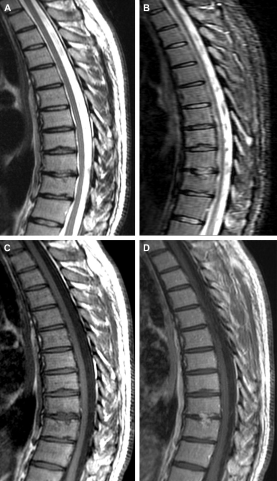
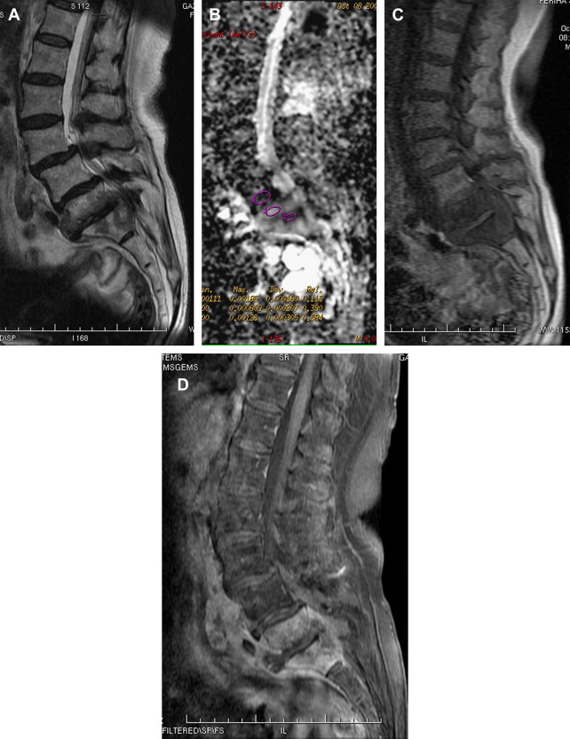
MR imaging is accepted as the gold standard in imaging spinal infections. With a reported sensitivity of 96%, specificity of 92%, and accuracy of 94%, MR imaging performs better than any other radiological technique or combined nuclear medicine studies. The major strength of MR imaging is its usefulness in early detection of infection when other modalities are still normal, such as radiographs, or nonspecific, as in nuclear medicine studies. For evaluating possible infection, MR imaging should cover the entire spinal axis and should include fat-suppressed T2-weighted imaging (T2WI) or short tau inversion recovery (STIR) images and fat-suppressed T1-weighted imaging (T1WI) to increase contrast enhancement conspicuity. Infection typically begins in the anterolateral part of the vertebral body near the end plate and then spreads to involve the intervertebral disk and neighboring vertebral body. The earliest sign of infection in adults is altered bone marrow signal, reflected as T2 hyperintensity, T1 hypointensity, and contrast enhancement, which is more pronounced along the end plates. Afterward, loss of definition of the end plate can be noticed. Findings of acute disk involvement are increased disk height and nonanatomic T2 hyperintensity. In the later stages of the diskitis, reduced disk height, loss of intranuclear cleft, and nonanatomic contrast enhancement are seen. Involvement of the normal disk may be missed due to its high signal on T2WI in cases without associated bony infection. Contrast administration is mandatory if there is a suspicion of infection ( Fig. 1 ). Infection can progress and cause loss of cortical continuity of the adjacent end plates and progressive destruction of vertebral body together with soft tissue infiltration ( Figs. 2 and 3 ), extending posteriorly into the epidural space and laterally into the paraspinal area. Articular processes and facet joints may be involved during the later stages. Reactive bone changes, new bone formation, sclerosis, kyphosis, and ankylosis are the late stage changes of the infectious process. In children, because the intervertebral disks can be directly inoculated, decreased disk height and contrast enhancement are the initial findings, followed by demineralization of adjacent vertebrae, end plate irregularities, and contiguous vertebral destruction. Overall findings yielding high sensitivity in diagnosing pyogenic spondylodiskitis are vertebral bone marrow edema, disk space T2 hyperintensity, disk enhancement, and epidural/paraspinal inflammation ( Box 1 ). In addition to these findings, pyogenic spondylodiskitis may present with atypical findings, such as lack of early bone marrow signal and end plate changes, involvement of a single or adjacent 2 vertebral bodies without the intervening disk, and discrete enhancing bony lesions mimicking metastasis. Combining clinical and laboratory information with MR imaging findings is important in solving those equivocal cases.


Invariably reduced disk height, non-anatomic T2 hyperintense, and enhancing disk
Irregularity, destruction, and enhancement of end plates and adjacent vertebral bodies
Epidural extension; phlegmon or abscess, or reactive enhancement
Soft tissue changes around spine: inhomogeneous paraspinal inflammatory swelling, abscess
Although MR imaging is accepted as the gold standard in imaging spine infection, it is not without challenges, such as in treatment response follow-up. It has been reported that MR imaging can lag behind the clinical picture with a delay of 4 to 8 weeks, even months after the initiation of antibiotherapy and disease response. Focal reinstitution of T1 signal hyperintensity in the bone marrow, reflecting bone marrow recovery with fatty infiltration, or decreased or absent contrast enhancement has been suggested as a marker of healing at MR imaging follow-up ( Box 2 , Fig. 4 ). Still, MR imaging is not a fully reliable technique in evaluating treatment response, especially in those patients who show clinical improvement on antibiotic treatment. Interpretation of MR imaging findings can also be challenging in the setting of postoperative spondylitis/spondylodiskitis. It has been reported that 2 parallel thin bands of enhancement in the disk space and/or paravertebral enhancement may favor spondylodiskitis over postoperative changes. Knowing that even uncomplicated postoperative spine can demonstrate disk or end plate signal changes together with enhancement, however, MR imaging cannot be used confidently in the differentiation of infection from postsurgical changes until at least 6 months after surgery. Pleural effusion also may be seen accompanying thoracal spondylitis. The possibility of spondylitis should be considered in the patients with pleural effusion of unknown cause. Pleural effusions may be sterile in majority of accompanying spondylodiskitis.


