KEY FACTS
Terminology
- •
Obstructed renal collecting system containing pus or infected urine
Imaging
- •
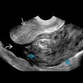
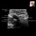
Presence of mobile debris and layering of low-amplitude echoes within dilated collecting system on US
- •
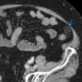
Dilated collecting system containing intermediate- or high-density material on CT
- •
Enlarged kidney with perinephric inflammatory changes on CT
Top Differential Diagnoses
- •
Sterile hydronephrosis
- •
Complex renal cyst
- •
Urothelial carcinoma
Pathology
- •
Stagnant urine becomes infected, filled with white blood cells, bacteria, debris, and pus
- •
Chronic > acute ureteral obstruction
- •
Etiology
- ○
Young adult: Calculus or ureteropelvic junction obstruction or duplicated collecting system more common
- ○
Elderly: Malignant ureteral stricture other mechanical obstruction
- ○
Clinical Issues
- •
Urologic emergency: Delay in diagnosis and treatment leads to irreversible renal parenchymal damage and renal failure
- ○
Progress to bacteremia or septic shock and can lead to 25-50% mortality
- ○
- •
Symptoms include fever, chills, flank pain
- •
Most common organism: Escherichia Coli
- •
Treatment: Percutaneous nephrostomy
- •
Diabetes is risk factor for worse clinical outcomes
- •
Early diagnosis and drainage are crucial to prevent bacteremia and septic shock
Scanning Tips
- •
Coronal plane best demonstrates collecting system, renal contour, renal cortex, medulla, and renal vessels
- •
To obtain good coronal view, place probe between iliac crest and lower costal margin; start with midaxillary window then try posterior axillary line where rib spaces are wider
- •
Graded probe pressure may help move bowel gas away
 in this case of pyonephrosis in an adult patient with multiple sclerosis on grayscale US imaging.
Related posts:


Stay updated, free articles. Join our Telegram channel

Full access? Get Clinical Tree


